BLOG
Amelogenesis Imperfecta: What It Is and How It Affects Teeth
Blog Tarihi: 14/06/2026

Understanding Amelogenesis Imperfecta (AI) in Clinical Practice
Amelogenesis imperfecta (AI) is a group of inherited conditions that disrupt normal enamel formation. For dental professionals, AI is more than an enamel “appearance issue”: it can change occlusal function, increase sensitivity, complicate adhesion, and influence long-term restorative planning. For patients, it often presents as teeth that look discolored or “worn down,” feel sensitive, and chip easily—sometimes from childhood.
This article is for educational purposes and aims to support clinical understanding and structured decision-making. Because AI varies widely between individuals and families, diagnosis and treatment planning should be individualized and based on comprehensive clinical and radiographic assessment.
What Exactly Is Amelogenesis Imperfecta?
AI refers to genetic enamel defects affecting primary and/or permanent dentitions, typically without an underlying systemic disease. The condition can be inherited in autosomal dominant, autosomal recessive, or X-linked patterns, and multiple genes involved in enamel matrix formation and mineralization have been implicated.
Clinically, AI is often categorized by the stage of enamel formation primarily affected:
- Hypoplastic AI: reduced enamel quantity (thin enamel, pits, grooves), often with relatively normal mineralization.
- Hypomaturation AI: enamel quantity may be near normal but enamel is softer and prone to chipping; opacities are common.
- Hypocalcified AI: enamel is poorly mineralized and can be very soft, with rapid wear and heavy staining.
These categories can overlap, and the phenotype may vary even within the same family. Recognizing the pattern is clinically useful because it informs adhesive choices, material selection, and prognosis.
How AI Affects Teeth: Common Signs and Patient Complaints
1) Enamel breakdown and accelerated wear
In many AI presentations—especially hypocalcified and hypomaturation forms—enamel may fracture under functional load. Patients can present with flattened occlusal surfaces, reduced clinical crown height, and visible dentin exposure.
2) Tooth sensitivity and discomfort
Thin or porous enamel can lead to thermal sensitivity. Dentin exposure due to enamel loss can amplify symptoms and affect oral hygiene habits, dietary choices, and overall quality of life.
3) Aesthetic concerns
Discoloration may range from chalky white opacities to yellow-brown staining. Surface irregularities, enamel pitting, and altered translucency can significantly impact smile aesthetics, especially in anterior teeth.
4) Functional and occlusal challenges
Severe wear may contribute to altered vertical dimension, occlusal instability, and parafunctional adaptation. When AI is identified early, preventive and interceptive measures may help reduce long-term structural loss.
5) Psychosocial impact
Although dental teams often focus on hard-tissue management, it is important to acknowledge that aesthetic changes and sensitivity can be socially limiting for patients—especially adolescents and young adults.
Diagnosis: Building a Reliable Clinical Picture
AI diagnosis is typically clinical, supported by radiographic findings and careful history. Key steps include:
- Medical and family history: similar findings among relatives can support inherited enamel pathology.
- Intraoral exam: note enamel thickness, surface texture, staining, and fracture patterns.
- Radiographic assessment: enamel may appear thin or have reduced radiopacity compared with dentin, depending on the type.
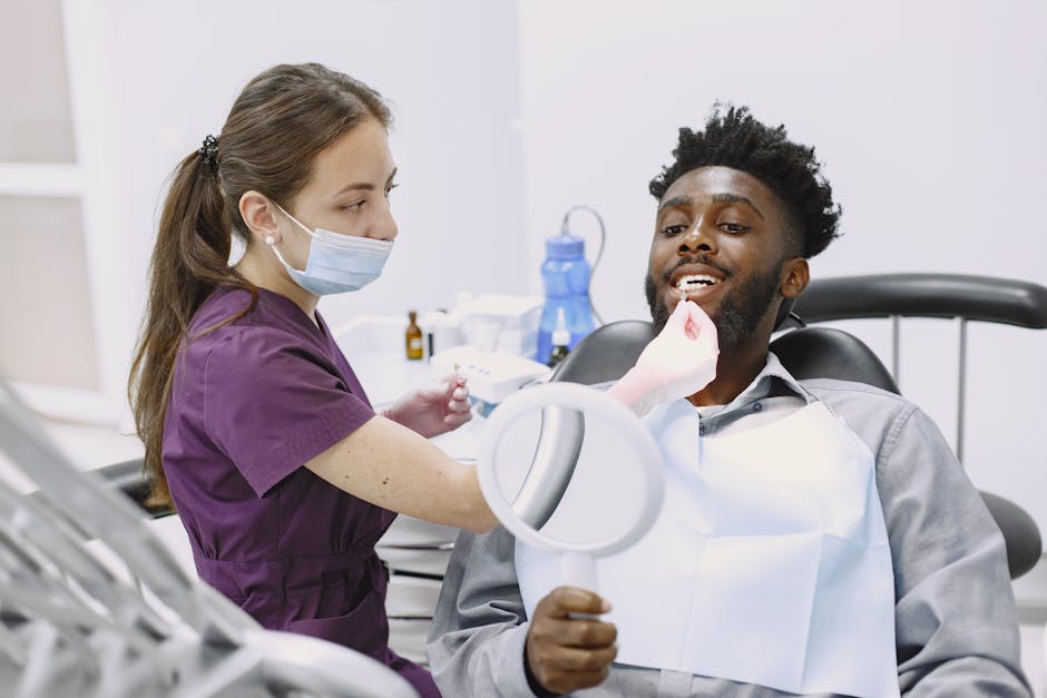
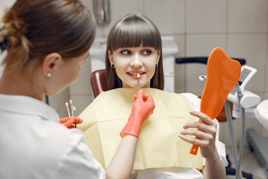
- Photographic documentation: standardized dental photography helps with baseline records, patient communication, and interdisciplinary planning.
Differential considerations can include fluorosis, molar-incisor hypomineralization (MIH), enamel hypoplasia from childhood illness, and erosion/attrition patterns unrelated to hereditary enamel defects. When presentation is complex, an interdisciplinary discussion can improve accuracy and planning.
Restorative Challenges in AI: Why Adhesion Is Not “Routine”
AI often complicates bonding because enamel may be thin, porous, or prism-deficient. Margins can end up on dentin more frequently, and the substrate can be variable across the same tooth. These factors can influence microleakage risk, marginal integrity, and restoration longevity.
For posterior teeth, clinicians frequently need a strategy that balances seal, stress management, and long-term maintainability. A practical refresher on isolation, selective etching, and liner/base considerations is highly relevant; see contemporary adhesive techniques for posterior restorations for a clinical-ready discussion that can be adapted to challenging enamel scenarios.
Clinical Pathways: From Prevention to Full-Mouth Rehabilitation
Management is case-dependent and may range from protective, minimally invasive strategies to comprehensive prosthodontic rehabilitation. The goal is typically to reduce sensitivity, preserve tooth structure, improve function, and address aesthetics—while maintaining realistic expectations regarding maintenance.
Early-stage and minimally invasive options
In mild cases or in younger patients, clinicians may prioritize prevention and protection:
- Desensitizing protocols and fluoride or remineralization adjuncts (as clinically appropriate).
- Sealants or conservative resin restorations to protect vulnerable surfaces.
- Occlusal guards when indicated for wear control.
Even when the definitive plan is prosthetic, incremental steps can stabilize the mouth and improve comfort while planning progresses.
Anterior aesthetics: When to consider porcelain laminate veneers
Patients with AI frequently request aesthetic improvements, but case selection is critical. Veneers may be considered when sufficient enamel is present for reliable bonding, when occlusion is favorable, and when expectations align with the limitations of compromised enamel substrates. In more severe AI, full-coverage options or hybrid approaches may be more predictable.
For clinicians refining indication criteria—such as enamel availability, parafunction risk, margin design, and minimally invasive preparation principles—review case selection for porcelain laminate veneers to support structured planning for AI-like presentations where enamel quality is uncertain.
Posterior protection and vertical dimension considerations
When posterior wear is advanced, restoring occlusal stability becomes a priority. Options may include indirect onlays/overlays, full-coverage crowns, or additive approaches depending on remaining tooth structure and functional demands. Digital workflows can assist in documenting the occlusal scheme, trialing vertical changes, and communicating treatment stages.
Endodontic Considerations: Sensitivity vs Pulpal Disease
AI-associated sensitivity does not automatically indicate pulpal pathology, but enamel loss and dentin exposure can increase risk of caries and pulpal inflammation. Deep restorations, cracks, or accelerated wear may lead to endodontic involvement in some cases.
When endodontic treatment is required, magnification and illumination can improve detection of calcified canals, cracks, or complex anatomy—especially in teeth compromised by long-term wear. For clinicians interested in ergonomics and precision, the dental operating microscope in modern endodontics outlines how enhanced vision may support more predictable workflows (education-focused, not a guarantee of outcomes).
When Teeth Become Non-Restorative: Extraction and Implant Pathways
In advanced AI cases, certain teeth may be structurally compromised to the point that restoration is not feasible. Treatment planning should be conservative and evidence-informed, but implant-supported rehabilitation can be part of the discussion when prognosis is poor.
For clinicians, timing and sequencing matter: if extraction is indicated, the approach may involve ridge preservation, immediate implant placement in selected cases, or staged protocols based on bone and soft-tissue conditions. A useful overview of decision points can be found in same-day tooth extraction and immediate implant placement, which reviews clinical considerations that may apply when managing compromised dentitions.
AI patients may also present with altered occlusal loads due to wear or parafunction, so prosthetic design and occlusal planning for implants should be particularly cautious and interdisciplinary.
Bone and Soft Tissue: Why Grafting Knowledge Matters
If long-standing tooth loss, infection, or traumatic extraction leads to ridge deficiencies, augmentation may be considered. Understanding grafting materials, membranes, staging, and risk management supports comprehensive planning—especially when aesthetics are a priority in the anterior zone.
For a focused educational update relevant to implant clinicians and residents, current approaches to bone grafting techniques in implant dentistry reviews contemporary concepts that may be used when ridge conditions are suboptimal.
Digital Dentistry and Dental Photography in AI Cases
AI cases often benefit from high-quality documentation and predictable communication. Digital tools can support:
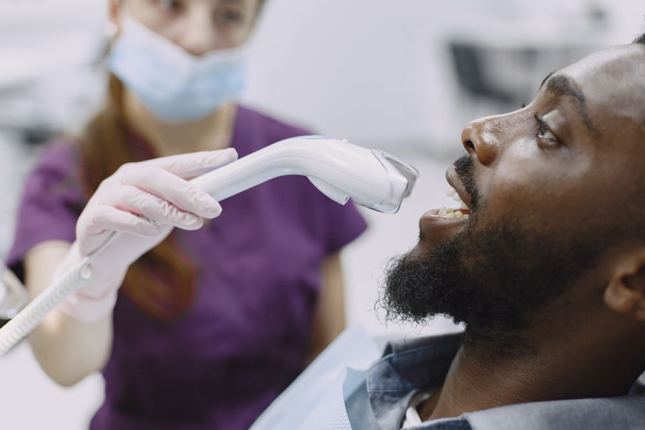
- Intraoral scanning to track wear progression, plan additive restorations, and reduce impression challenges in sensitive patients.
- Digital smile design to align expectations, explore minimal vs comprehensive options, and facilitate lab communication.
- Dental photography for baseline records, shade mapping, and patient education.
In a teaching environment, these tools also make treatment planning more transparent for trainees—linking diagnosis to material selection and long-term maintenance protocols.
Learning Focus: Turning Complex AI Cases into Structured Clinical Training
For dentists and dental students, AI is a valuable “framework condition” because it forces careful thinking about adhesion, occlusion, aesthetics, and interdisciplinary sequencing. It also highlights why a single-material approach rarely works: the substrate can change from tooth to tooth, and the plan often evolves through phases.
At Istanbul Dental Academy, our continuing dental education philosophy emphasizes clinical reasoning plus hands-on repetition—whether the topic is adhesive restorative strategies, aesthetic dentistry workflows, endodontic magnification, or implant-prosthetic planning. Complex enamel cases like AI are excellent learning opportunities to practice documentation, rubber dam protocols, mock-ups, provisionalization strategies, and digital planning under mentorship.
Key Takeaways
- Amelogenesis imperfecta is an inherited enamel disorder with variable clinical presentations and restorative implications.
- Common impacts include enamel breakdown, sensitivity, aesthetic concerns, and occlusal wear—often requiring staged, interdisciplinary care.
- Adhesion can be unpredictable in AI; material choice and technique sensitivity are central to long-term success.
- In advanced cases, endodontics, prosthodontics, and implant dentistry (including grafting) may be part of the treatment pathway.
This content is for educational purposes and does not replace individualized diagnosis or treatment planning by a licensed dental professional.
Diğer Yazılar


