BLOG
Digital Impression Techniques for Implant-Supported Prostheses
Blog Tarihi: 25/06/2026
Why digital impressions matter in implant prosthodontics
In implant-supported prostheses, precision is not a luxury—it is a prerequisite. The impression phase determines how accurately the implant positions are transferred to the laboratory, directly influencing passive fit, occlusal stability, screw mechanics, and long-term maintenance. In recent years, digital impression systems (intraoral scanners + scan bodies + CAD/CAM workflows) have become mainstream in clinics across Istanbul and internationally, changing how dentists approach implant prosthodontics.
Digital workflows can streamline appointments, improve patient experience, and simplify communication with the dental laboratory. Yet clinical predictability depends on understanding scanner limitations, scan body handling, soft-tissue management, and how to verify the record—especially in complex cases such as multiple implants or full-arch restorations. This article is for educational purposes and is intended to support clinicians and dental students who want a structured, clinically oriented overview of digital impression techniques for implant-supported restorations.
From conventional to digital: what actually changes?
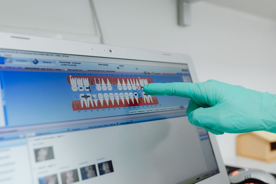
Conventional implant impressions rely on impression copings, open- or closed-tray techniques, impression materials, and analogs. Digital impressions replace the physical impression material with a sequence of optical images that are stitched into a 3D mesh. Implant position is captured through a scan body (or scan post) that is recognized by CAD software via its geometry.
The practical shift is not merely “taking a scan instead of an impression.” It changes:
1) Error sources: polymerization shrinkage and tray distortion are replaced by scan stitching errors, reflective surfaces, and scan body instability.
2) Soft-tissue strategy: the emergence profile and peri-implant mucosa are often recorded through scanning rather than through customized impression copings and gingival masks.
3) Verification methods: clinicians may use digital cross-arch measurements, bite scans, and (in some workflows) photogrammetry or verification jigs.
Core components of a digital implant impression workflow
Intraoral scanner performance and calibration
Not all scanners behave the same in implant cases. Factors such as scanning technology, software algorithms, and “best-fit” stitching approaches can influence accuracy—especially over long spans. Regular calibration (as recommended by the manufacturer) and consistent scanning protocols help reduce variability.
Scan bodies: selection, seating, and stability
Scan bodies act as the “transfer device” between the mouth and CAD software. Small seating errors can translate into prosthetic misfit. Clinical takeaways include:
Confirm full seating: remove soft tissue or debris that prevents complete seating; verify with direct vision and, when needed, radiographic confirmation.
Control torque and handling: follow manufacturer torque guidelines; over-tightening can deform components, under-tightening can cause micromovement during scanning.
Keep surfaces clean and dry: saliva, blood, or powder residue can disrupt scanner recognition, especially for glossy scan bodies.
Soft-tissue management around implants
Digital accuracy is influenced by how well the peri-implant tissues are controlled during scanning. Good retraction and moisture control improve capture of the emergence contour and margins—particularly in implant-supported crowns with customized abutments.
Clinicians interested in advanced peri-implant soft-tissue concepts may also explore materials used for tissue augmentation. For example, acellular dermal matrices have become part of the broader conversation around tissue volume and contour management in implant cases; see acellular dermal matrix use in dentistry for a focused educational overview.
Clinical scan strategies: single-unit vs. multiple implants
Single implant (posterior or anterior)
Single-unit cases are often the easiest entry point into digital implant impressions. A typical strategy is to capture: opposing arch, working arch with scan body, and a bite scan. Clinically, keep scan paths short and stable, and avoid “wandering” across reflective restorative materials unnecessarily.
In the esthetic zone, clinicians may integrate digital dentistry principles with smile design planning. A stable digital impression becomes the foundation for designing the emergence profile, crown contours, and occlusal scheme—often alongside clinical photography and shade documentation.
Multiple implants (short-span bridges)
As the inter-implant distance increases, the cumulative risk of stitching errors can rise. Practical tips include:
Segmented scanning: scan key areas first (scan bodies and adjacent teeth), then fill in soft tissues.
Use stable landmarks: teeth provide strong reference geometry; edentulous areas may require careful scanning to avoid “featureless” regions.
Check scan body readability: ensure all scan bodies are clearly captured from multiple angles for reliable software recognition.
Full-arch implant prostheses
Full-arch digital impressions are clinically feasible, but technique sensitivity increases significantly. Long-span accuracy depends on scanner capabilities, scan strategy, the presence (or absence) of teeth, and soft-tissue mobility. In some protocols, clinicians consider auxiliary methods (e.g., reference markers, scan appliances, or photogrammetry) to enhance reliability.
When planning full-arch implant cases, systemic health and maintenance risk should be part of the broader clinical evaluation. For clinicians treating medically complex patients, Istanbul Dental Academy encourages structured learning around risk assessment and communication. Relevant reading includes Dental implants for patients with diabetes: what clinicians should know and diabetes and tooth loss: implant planning and clinical considerations, which discuss educational considerations for treatment planning and follow-up.
Occlusion and the bite record: the often underestimated step
Even a perfect implant scan can be compromised by an inaccurate bite registration. Digital bite scans may be affected by limited interocclusal space, patient movement, scanning too small an area, or scanning at an unstable mandibular position. Clinicians can improve predictability by:
Capturing a broad bite area: include multiple posterior contacts when possible.
Confirming seating of scan bodies and abutments: avoid changes between working scan and bite scan.
Verifying occlusion clinically: use shimstock/articulating paper at try-in; consider a second verification if the patient reports a different bite sensation.
Soft-tissue and saliva: practical implications for digital scanning
Moisture control is central to optical scanning. Excess saliva can create reflective pooling, blur scan body edges, and reduce capture of mucosal details. Conversely, xerostomia (low salivary flow) can affect comfort and tissue behavior, and may be relevant to halitosis risk and prosthesis tolerance.
For a broader clinical discussion, see natural ways to increase saliva flow: a clinical guide for dental professionals and halitosis and saliva: the clinical link every dentist should know. While these topics may appear separate from digital impressions, they influence chairside scanning conditions, patient experience, and ongoing maintenance around implant restorations.
Common pitfalls and how to reduce them
1) Incomplete scan body capture
If the scan body is missing critical surfaces or edges, the CAD library may align it incorrectly. Re-scan from multiple angles and ensure adequate retraction. Avoid “quick scans” that capture only the top surface.
2) Stitching errors across edentulous spans
Edentulous tissue can be mobile and uniform, making alignment harder. Use a consistent scan path and return frequently to stable reference areas (teeth, scan bodies). Consider clinical protocols that add reference geometry when appropriate.
3) Tissue collapse at the emergence profile
After removing a provisional or healing abutment, soft tissue can change quickly. Efficient workflow matters: prepare for scanning by having scan bodies ready, use gentle retraction, and scan promptly. In highly esthetic cases, clinicians may use customized provisionals or healing components to shape tissue before final scanning.
4) Overreliance on the “digital” label
Digital impressions are not automatically more accurate in every scenario. A clinician’s technique, case selection, and verification steps still determine success. A balanced approach—knowing when digital is ideal and when conventional or hybrid methods may be safer—reflects mature clinical decision-making.
Lab communication: turning scans into restorations
Digital implant prosthodontics shines when the clinic and laboratory communicate effectively. Alongside the scan files, consider sending:
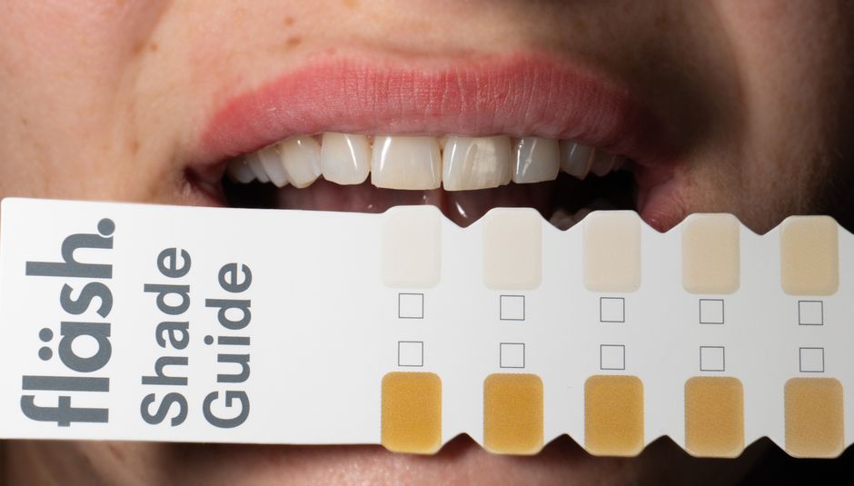
High-quality photographs: frontal, lateral, occlusal, retracted views; shade tabs; stump shade (for customized abutments).
Design notes: occlusal scheme, proximal contact preferences, emergence profile goals, and screw-access considerations.
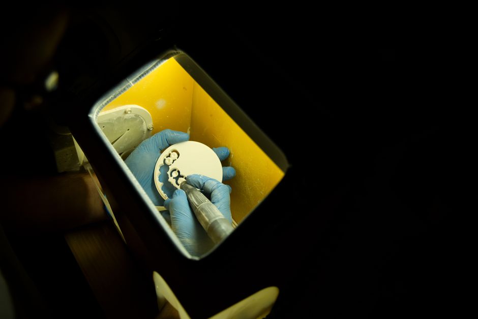
Material selection rationale: zirconia, titanium-base solutions, hybrid ceramics, or other systems depending on load and esthetics.
Dental photography and documentation are increasingly integrated into digital prosthodontic workflows, supporting more predictable esthetic outcomes and fewer remakes.
How Istanbul Dental Academy approaches training in digital implant impressions
Clinicians often find that the biggest leap is not buying a scanner—it is building a repeatable clinical protocol. At Istanbul Dental Academy, our continuing dental education philosophy emphasizes hands-on training and step-by-step workflows that connect diagnosis, implant surgery/prosthetics, scanning technique, and restoration delivery.
In practical sessions, participants typically focus on topics such as scan body handling, soft-tissue control, scanning strategies for different arch conditions, and how to evaluate scan quality before sending to the lab. These competencies complement broader implant education, restorative dentistry, prosthodontics, and digital dentistry learning paths—helping dentists translate technology into daily clinical outcomes.
Key takeaways
Digital impression techniques for implant-supported prostheses can enhance efficiency and communication, but accuracy depends on clinical fundamentals: scan body seating, tissue control, scan strategy, and bite verification. As implant cases become more complex—multiple implants, full arches, medically nuanced patients—the value of structured training and protocol-based execution increases.
This content is for educational purposes and does not replace individualized clinical judgment, diagnosis, or manufacturer-specific guidance. For dentists looking to strengthen their digital implant workflow with hands-on practice, Istanbul Dental Academy’s courses provide an opportunity to refine techniques in a supervised learning environment.
Diğer Yazılar