BLOG
Advantages of Using a Dental Microscope in Endodontics
Blog Tarihi: 25/06/2026

Why the Dental Microscope Has Become Central to Modern Endodontics
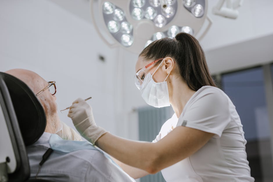
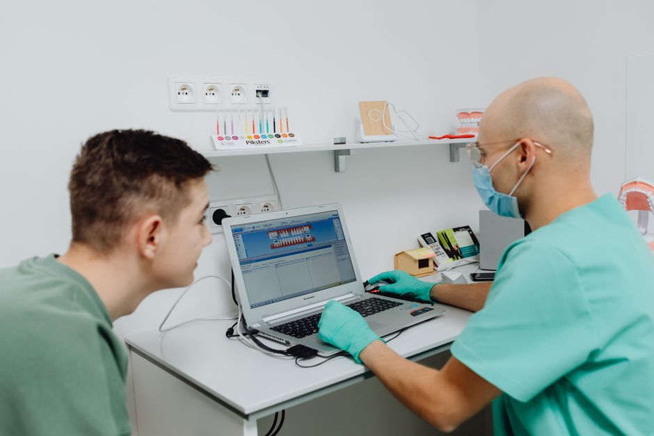
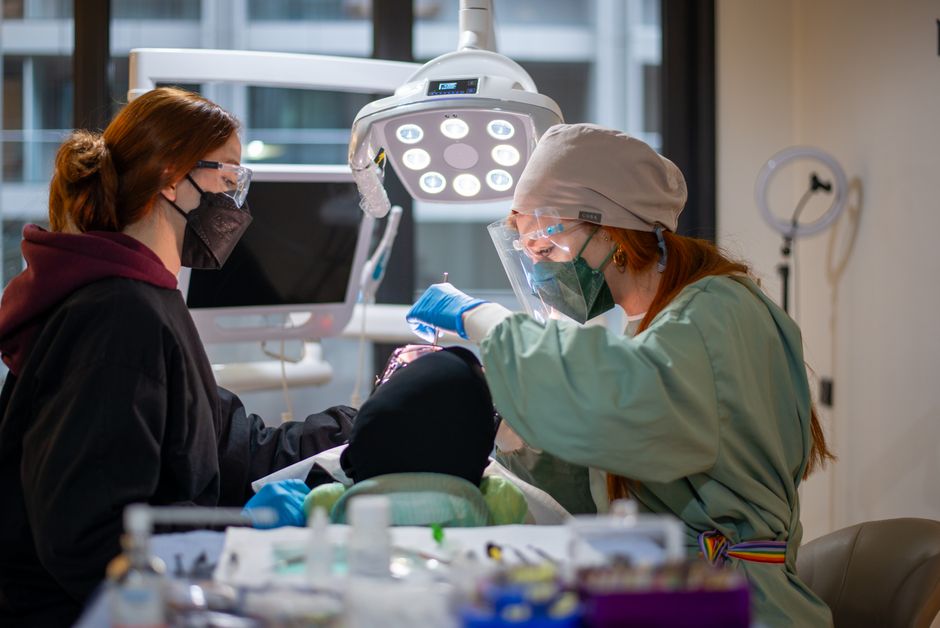
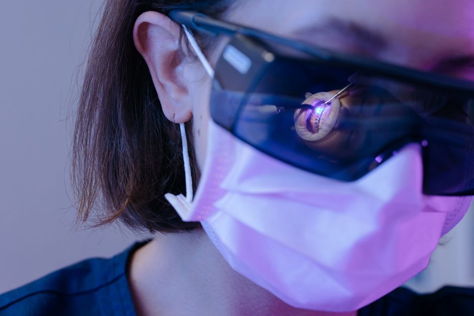
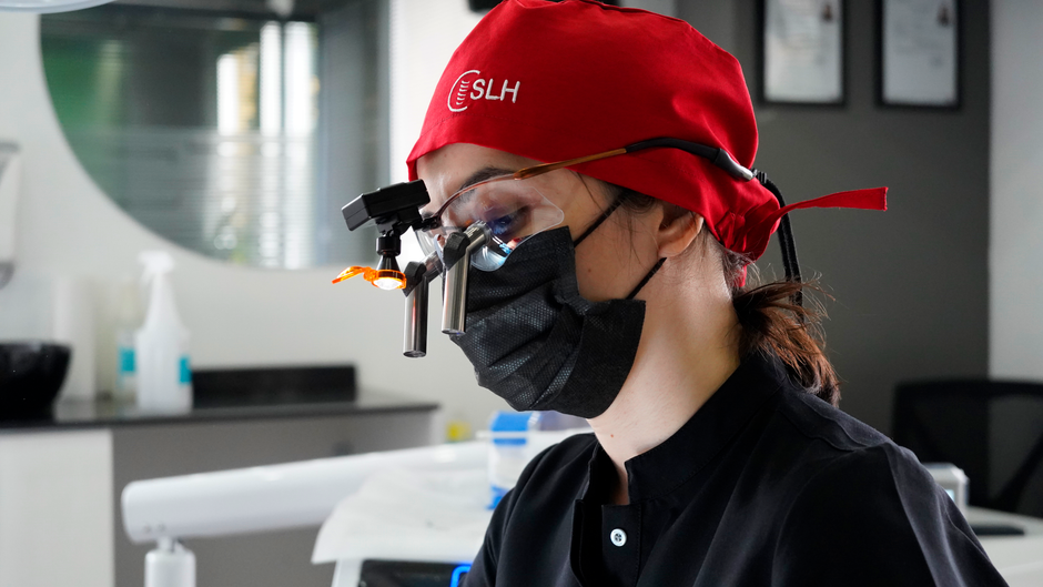
Endodontics is often decided in millimeters—and sometimes fractions of a millimeter. Locating a calcified canal, identifying an isthmus, negotiating a separated instrument, or confirming a crack line can hinge on whether the clinician can truly see what is happening inside the tooth. For many dental professionals, the dental operating microscope has shifted from being a “nice-to-have” to a core tool for improving visualization, supporting minimally invasive access, and building confidence in complex cases.
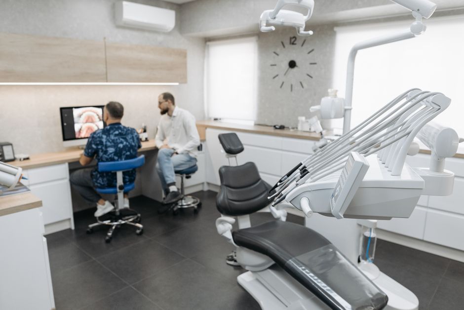
In Istanbul, where patients frequently expect both high-quality outcomes and efficient appointments, clinicians also face increasing pressure to make accurate, well-documented decisions. Microscope-assisted endodontics can help bridge that gap—especially when combined with contemporary imaging, rubber dam isolation, ultrasonics, and high-quality adhesive and restorative protocols. This content is for educational purposes and is not a substitute for individualized clinical judgment or patient-specific care planning.
1) Enhanced Visualization: Seeing More to Do Less
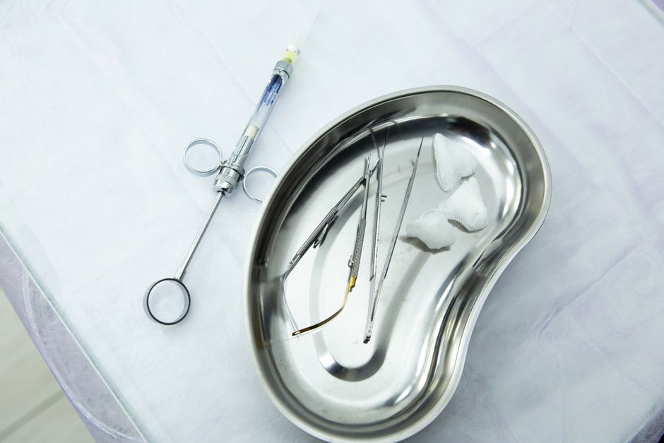
The most obvious advantage of a dental microscope is magnification paired with coaxial illumination. Unlike traditional overhead lighting, microscope illumination aligns with the clinician’s line of sight, reducing shadows in deep cavities and allowing finer evaluation of internal anatomy. This matters across the entire endodontic pathway:
During diagnosis, magnification can support the inspection of fracture lines, coronal leakage, restoration margins, and pulp chamber floor anatomy. During access, improved visualization may allow more conservative cavity design and more precise troughing. During shaping and disinfection, clinicians can better evaluate chamber cleanliness and detect anatomical complexities that could compromise irrigation efficacy.
Microscope use is also increasingly relevant in restorative planning. If endodontic access is conservative and margins are clean, subsequent adhesive restoration may be more predictable. For clinicians refining their coronal sealing approach after root canal treatment, consider aligning microscope-assisted access refinement with evidence-based bonding workflows, such as those discussed in Contemporary Adhesive Techniques for Posterior Restorations: A Clinical-Ready Guide.
2) Improved Canal Location and Management of Complex Anatomy
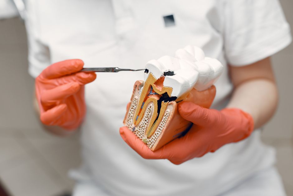
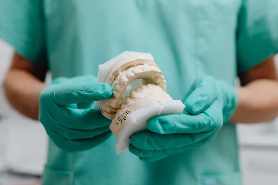
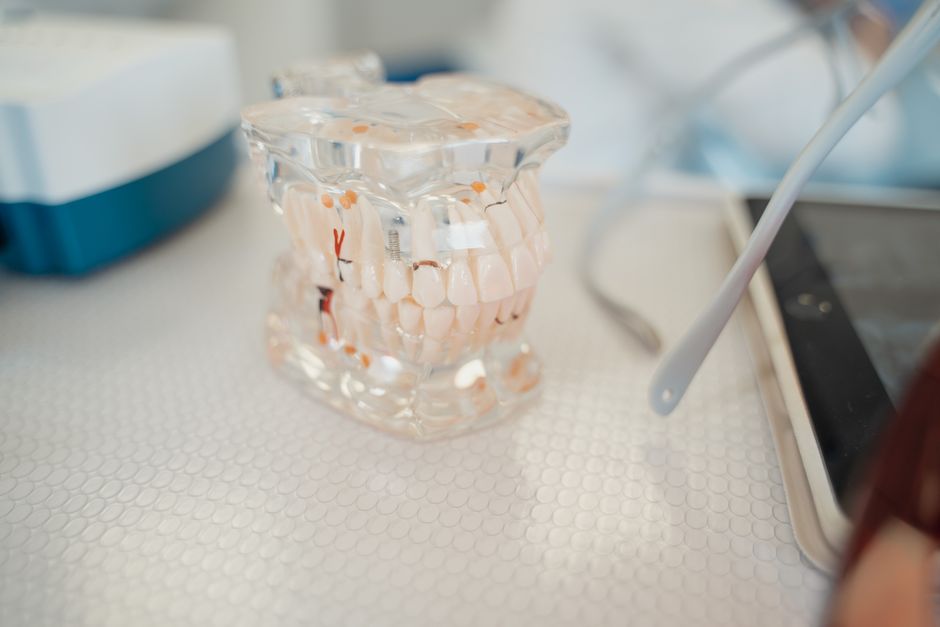
Missed canals remain a classic reason for persistent apical pathology and retreatment. Maxillary molar MB2 canals, mandibular incisors with two canals, C-shaped mandibular second molars, and calcified systems are scenarios where magnification can meaningfully influence detection and negotiation.
Calcified canals and conservative troughing
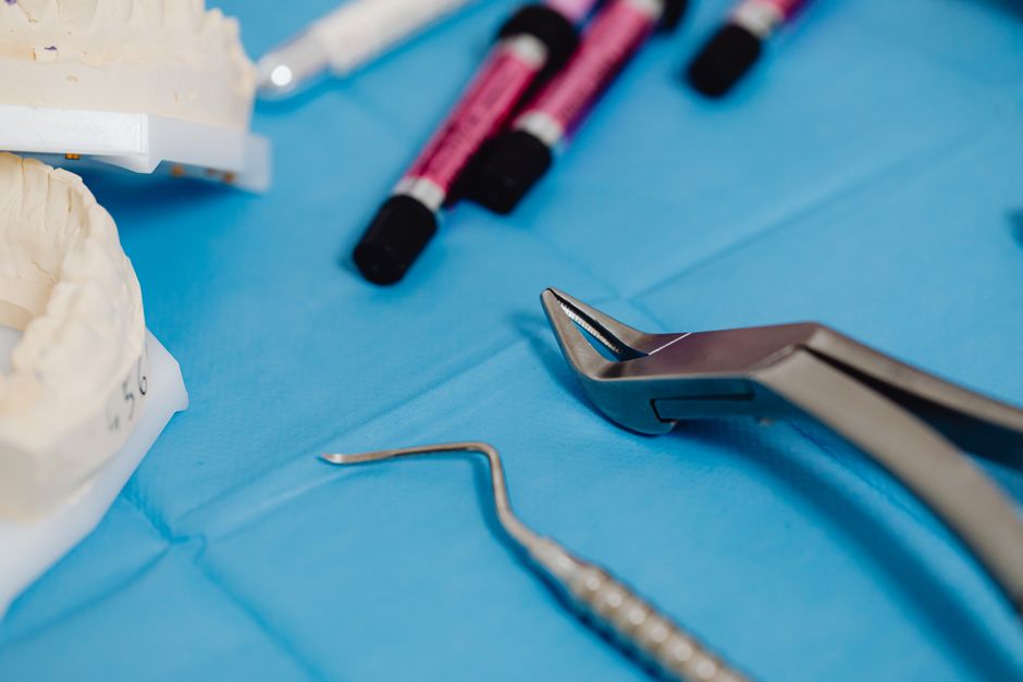
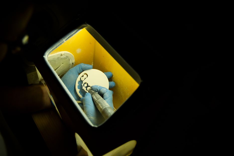
In calcified cases, the microscope can support a conservative approach by enabling controlled dentin removal. Combined with ultrasonics, troughing under magnification may reduce the risk of excessive dentin sacrifice or perforation—particularly in teeth with thin walls or previous restorations. While magnification does not replace sound anatomical knowledge, it can make anatomical landmarks easier to interpret and may help clinicians stay oriented to the pulp chamber floor map.
Isthmuses and fins: anatomy that shapes outcome expectations
Magnification may also help clinicians recognize isthmuses or fins that are difficult to instrument mechanically and rely more on irrigation protocols. In educational settings, discussing these “unreachable” spaces can be a turning point: the microscope makes complexity visible, which can lead to more realistic risk communication, improved disinfection strategy selection, and better restorative sealing decisions.
3) Microscope-Assisted Retreatment: A More Controlled Workflow
Retreatment is often where the microscope shows its most practical value. Removing old filling materials, locating missed anatomy, addressing ledges, and managing procedural complications typically demands precision, patience, and reliable illumination. Under magnification, clinicians may feel better able to:
Identify the entry point to a missed canal, especially when obscured by previous obturation or restorative material.
Use ultrasonics more safely for troughing and material removal while monitoring dentin thickness.
Assess the chamber for hidden cracks, perforations, or unusual anatomy that can change prognosis discussions.
Microscope use may also support better interdisciplinary decision-making. Some retreatment cases are ultimately restored with indirect restorations or integrated into broader aesthetic plans. Understanding how endodontic stability interacts with cosmetic dentistry can guide case selection—particularly in high-demand smile cases. For a clinical perspective on selecting restorative paths, you may find it helpful to review Hollywood Smile vs Zirconia Crowns: Clinical Differences and Case Selection, which frames how restorative choices can align with functional and aesthetic goals.
4) Better Management of Iatrogenic Events (and More Transparent Risk Assessment)
Even experienced clinicians encounter complications. A microscope does not prevent every problem, but it may improve the clinician’s ability to assess and manage certain events with more control and documentation.
Separated instruments
Magnification can help identify the fragment location and assess whether bypassing or removal is feasible without excessive dentin removal. This supports more informed decision-making, including when referral is the best option.
Perforations
Early detection can be critical. Under magnification, perforation sites may be easier to visualize, debride, and manage with modern repair materials—while keeping a clear view of margins and bleeding control. The microscope can also help clinicians judge whether a perforation is restorable and periodontally maintainable, supporting more realistic patient communication.
Cracks and structural integrity
Crack identification is nuanced, and magnification can improve inspection of the pulpal floor and internal walls. However, diagnosing vertical root fracture remains complex and typically involves correlation with symptoms, radiographs/CBCT, and periodontal findings. The microscope can contribute to the overall diagnostic picture rather than serving as a single definitive tool.
5) Ergonomics and Longevity: A “Hidden” Advantage
Endodontics can be physically demanding: neck flexion, shoulder tension, and prolonged static posture are common contributors to clinician fatigue. A microscope can support a more upright posture, potentially reducing musculoskeletal strain over years of clinical practice. Many dentists report that when posture improves, fine motor control and endurance during complex cases also improve.
That said, ergonomics depend on correct microscope positioning, operator seating, and assistant coordination. This is where structured training becomes essential: the benefits of magnification are often limited if the clinician struggles with posture, focus, or hand stability under high magnification.
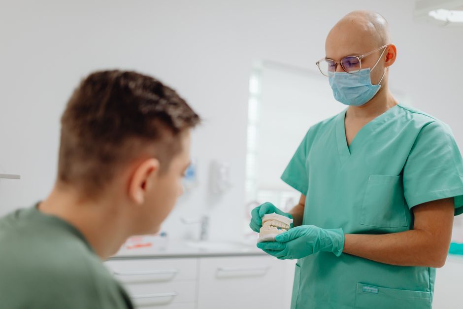
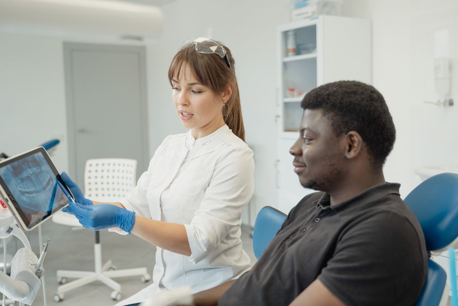
6) Documentation, Communication, and the Patient Trust Factor
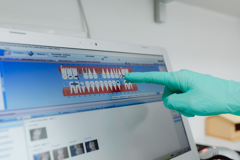
Modern microscopes frequently integrate photo and video capture. For dental professionals, documentation can support:
Case presentations and patient education (e.g., showing a hidden canal or a crack line).
Interdisciplinary referrals by sharing clear visuals with prosthodontists, periodontists, or oral surgeons.
Self-audit and learning, especially when reviewing outcomes and refining technique over time.
When combined with dental photography principles—composition, lighting, and consistent capture protocols—microscope documentation can elevate clinical communication. It can also support ethical informed consent discussions by making findings more tangible to patients.
7) Integration with Digital Dentistry and Implant Treatment Planning
Microscope-assisted endodontics doesn’t exist in isolation. In contemporary practice, clinicians commonly evaluate whether to preserve a tooth endodontically, restore it, or replace it with an implant-supported solution. High-quality visualization can improve the assessment of restorable tooth structure, ferrule potential, and crack extent—factors that often influence the “save vs. replace” conversation.
For example, if endodontic prognosis is uncertain due to structural compromise, clinicians may consider implant alternatives depending on systemic risk factors, bone conditions, and patient expectations. If you’re exploring how implant workflows are evolving for efficiency and predictability, you may be interested in Is One-Day Dental Implant Treatment Really Possible? as well as Digital Planning for Full-Arch Implant Cases: A Modern Workflow for Predictable Outcomes. Understanding these pathways helps clinicians frame endodontic treatment as part of a broader rehabilitation plan rather than a standalone procedure.
8) Periodontal and Soft-Tissue Considerations Around Endodontically Treated Teeth
Endodontic success is tied to periodontal health and restorability. Even when the root canal procedure is technically sound, soft-tissue management and coronal seal quality influence long-term stability. In certain cases—particularly where recession, thin phenotype, or restorative margin management is relevant—soft-tissue augmentation concepts may enter the conversation during comprehensive planning.
Materials used in periodontal plastic procedures are outside endodontics per se, but understanding them supports interdisciplinary communication. For an educational overview of one such biomaterial, see Asellüler Dermal Matriks: Diş Hekimliğinde Kullanımı. Seeing the “full picture” helps clinicians plan restorations that respect soft tissue and support maintainability—especially in aesthetic zones.
Common Learning Curves with Microscope Dentistry (and How Training Helps)
Clinicians often discover that the microscope is not simply a piece of equipment—it’s a change in operating style. Typical learning curves include:
Hand-eye coordination at higher magnification: Small movements look larger, and fine motor control becomes more noticeable.
Assistant choreography: Four-handed dentistry under a microscope requires coordinated suction, mirror use, and instrument transfer.
Field isolation: Rubber dam placement, clamp stability, and moisture control become even more critical when the visual field is magnified.
Time management: Early microscope users may experience longer appointments until workflow becomes familiar.
At Istanbul Dental Academy, many participants seek continuing dental education that bridges theory and clinical reality. Hands-on courses—especially those that combine magnification, ultrasonics, modern obturation strategies, and post-endodontic adhesive restoration concepts—can help dentists integrate microscope workflows more confidently into daily practice. The goal is not to treat the microscope as a “magic solution,” but as a tool that enhances precision when paired with sound clinical protocols.
Practical Takeaways for Dentists Considering a Dental Microscope
Choose indications strategically. Start with cases where the microscope’s advantage is immediate—canal location in molars, retreatment access refinement, or conservative access in teeth with significant restorative demands.
Build a repeatable setup. Consistent patient positioning, operator posture, and microscope alignment reduce fatigue and shorten the learning curve.
Integrate restorative planning early. Think beyond obturation: the quality of the coronal seal and restoration design are essential to long-term outcomes.
Document and reflect. Photos and videos can support patient communication, interdisciplinary collaboration, and personal improvement.
Ultimately, microscope-assisted endodontics reflects a broader trend in dentistry: higher precision, better documentation, and more interdisciplinary planning. With the right training and clinical workflows, magnification can help clinicians approach complex endodontic scenarios with greater clarity and control. This content is for educational purposes and does not replace professional judgment, local regulations, or patient-specific assessment.
Diğer Yazılar