
BLOG
Managing and Preventing Sinus Perforations in Implant and Oral Surgery
Blog Tarihi: 15/09/2026
Sinus perforations: why this topic matters in daily practice
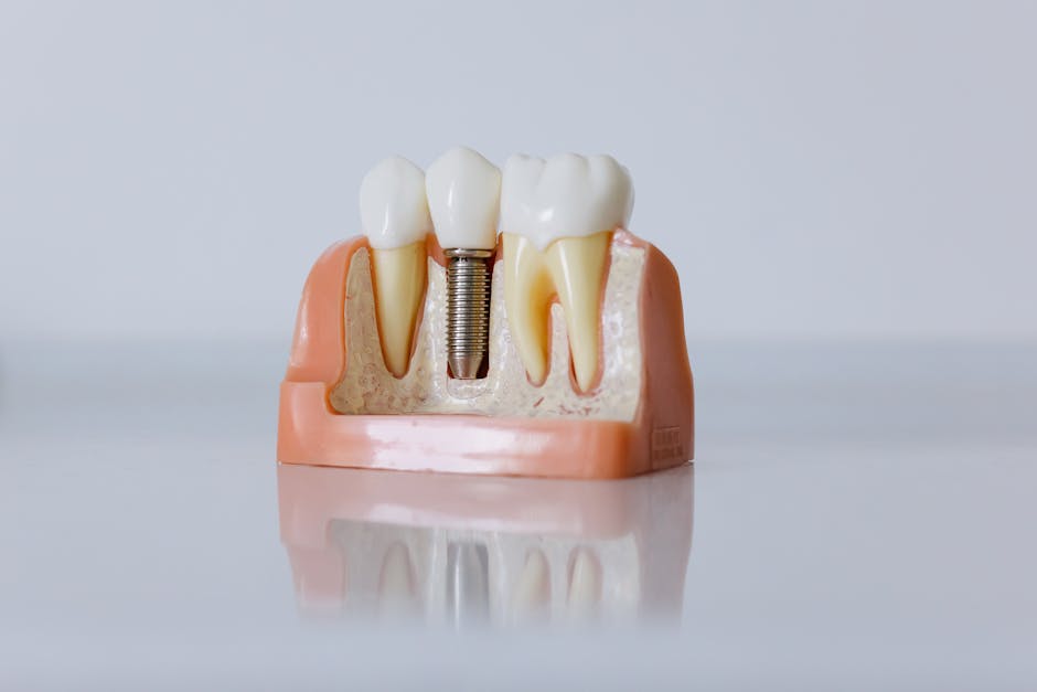
In the posterior maxilla, the anatomical relationship between the alveolar ridge and the maxillary sinus makes many procedures—implant site preparation, extractions, sinus augmentation, and even endodontic retreatment—technically sensitive. A sinus membrane perforation (commonly referring to Schneiderian membrane tears during sinus elevation) or an oroantral communication (OAC) after extraction can complicate healing, compromise graft stability, and increase the risk of sinusitis. For clinicians, the goal is not only to “manage” perforations when they happen, but to prevent them through structured planning and refined technique.
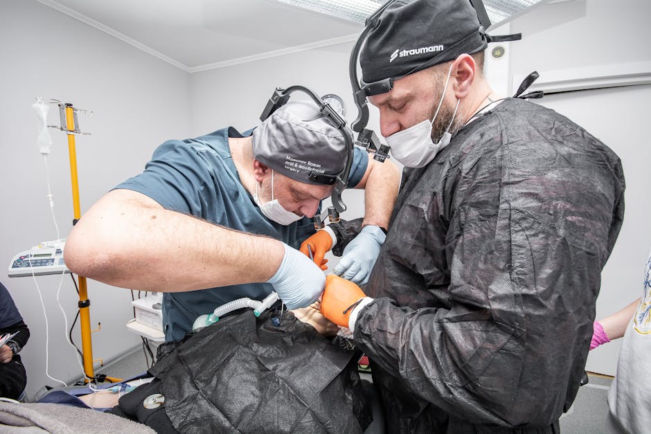
This article is for educational purposes and is designed for dentists, dental students, and clinicians pursuing continuing dental education (CDE). At Istanbul Dental Academy, these topics are often integrated into hands-on training pathways—especially in implant dentistry and oral surgery—where anatomy, imaging interpretation, flap design, and complication protocols are taught as a complete clinical workflow.
Terminology: perforation, OAC, and OAF
“Sinus perforation” is used broadly in clinical conversations, but it is helpful to differentiate:
Schneiderian membrane perforation: A tear of the sinus lining, most often during lateral window or transcrestal sinus elevation. The primary concern is graft migration, reduced regenerative stability, and postoperative sinus inflammation.
Oroantral communication (OAC): A direct opening between the oral cavity and the maxillary sinus, frequently after extraction of maxillary molars/premolars, removal of cystic lesions, or implant-related complications.
Oroantral fistula (OAF): An epithelialized, persistent tract that develops when an OAC fails to close, typically becoming a chronic problem requiring surgical management.
Risk factors: when perforations are more likely
Anatomical and radiographic factors
Posterior maxillary anatomy varies widely. Common risk enhancers include limited residual bone height, sinus septa, thin Schneiderian membrane, narrow palatal vault, and pneumatized sinus with irregular floor morphology. Periapical pathology, previous extractions, or iatrogenic defects may further reduce the predictability of sinus-related procedures.
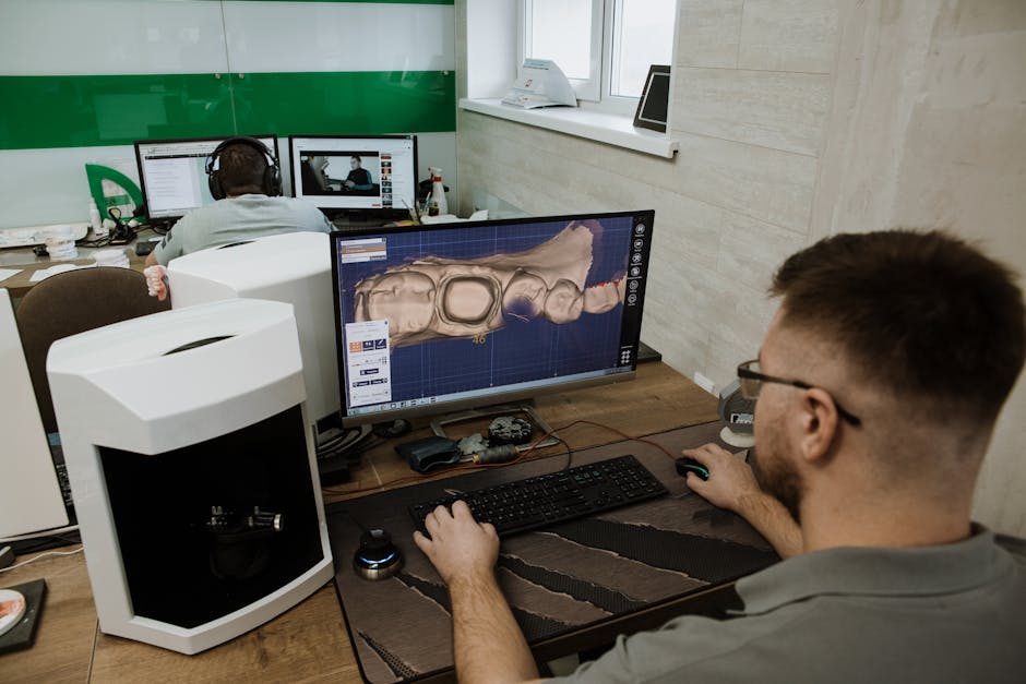
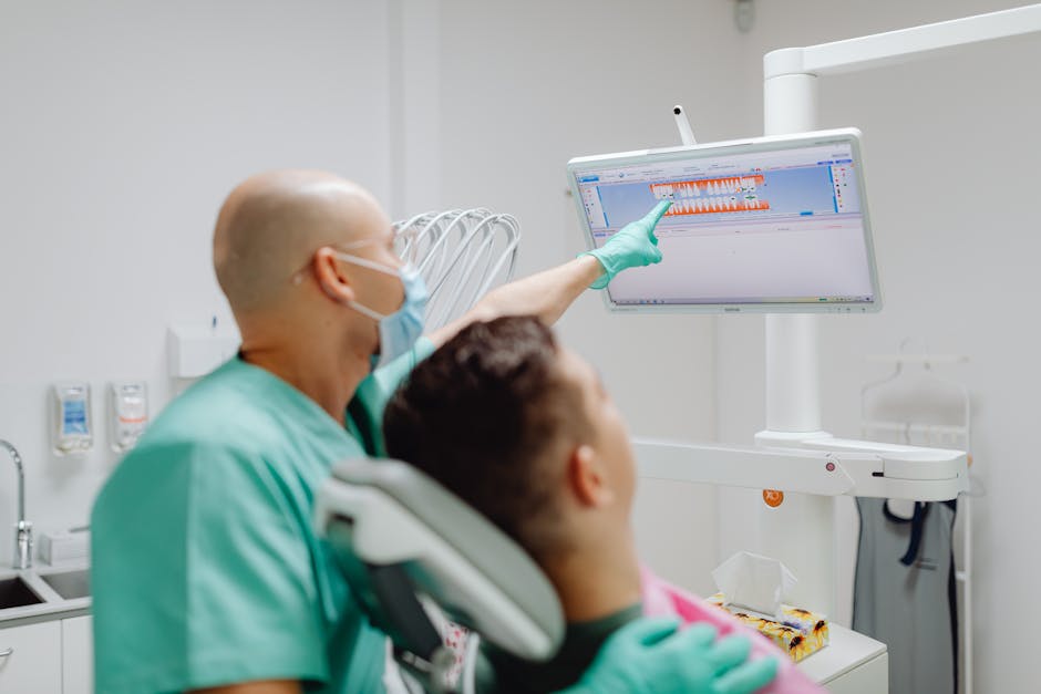
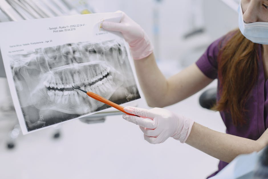
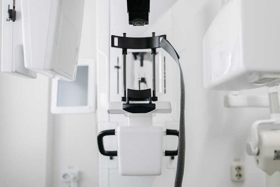
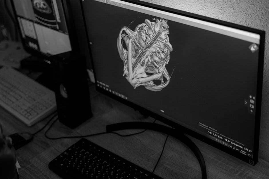
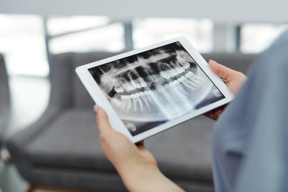
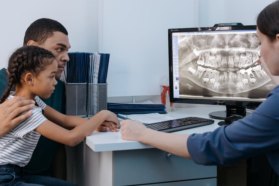
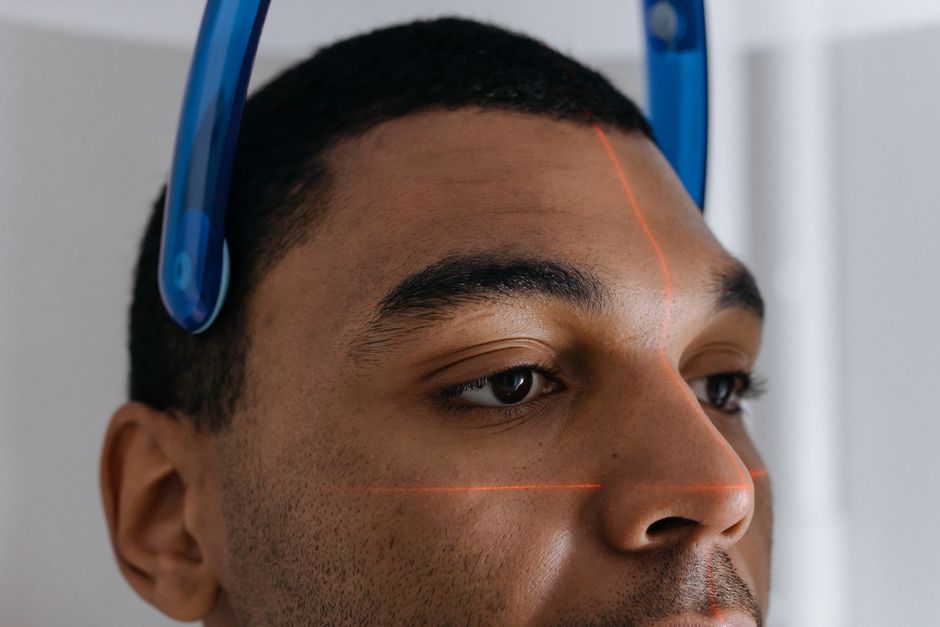
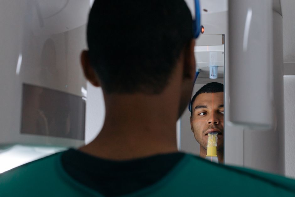
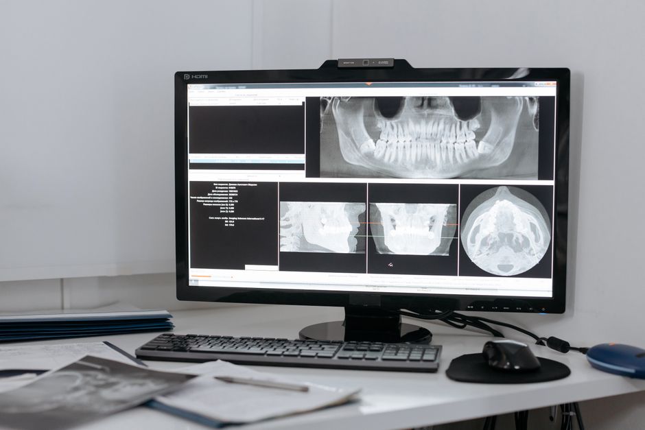
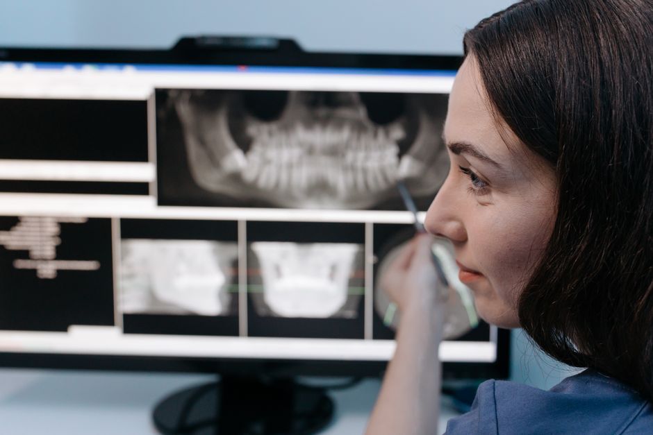
Advanced imaging is a key preventive tool. Clinicians planning implants in the posterior maxilla often rely on CBCT to visualize residual ridge height, sinus septa, ostium proximity, membrane thickness, and the presence of mucosal thickening or pathology. For a clinical workflow perspective, see Why CBCT Matters in Dental Implant Planning: A Clinical, Digital Workflow Guide, which aligns well with how modern implant planning is taught in digital dentistry modules.
Procedure-related factors
Perforations are more common with aggressive instrumentation, poorly positioned lateral windows, uncontrolled elevation forces, and inadequate flap access. In transcrestal approaches, excessive osteotome force or misjudged drilling depth can compromise the membrane. In extraction cases, excessive apical pressure or levering can create an OAC, particularly when the sinus floor is thin and root anatomy is divergent.
Prevention: a structured approach clinicians can apply
1) Start with a diagnostic workflow, not a “procedure plan”
Effective prevention begins by treating diagnosis and risk assessment as a workflow. Evaluate systemic factors (smoking, uncontrolled diabetes, chronic sinusitis history), local factors (periodontal support, keratinized tissue, biotype), and imaging findings. When sinus health is uncertain, collaboration with ENT specialists may be considered, especially for symptomatic or radiographically suspicious cases. This is not about delaying care—it is about improving predictability.
2) Use digital planning to reduce surprises
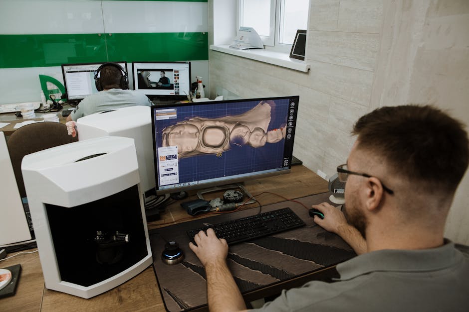
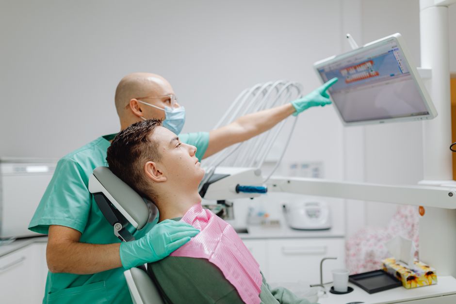
In education settings, the most repeatable reduction in complications often comes from improved imaging interpretation and guided planning. Surgical guides and digitally planned implant positions can help control angulation and depth, which is particularly relevant near the sinus floor. These concepts are reinforced in hands-on implant training where participants learn how to translate CBCT findings into safe drilling protocols.
3) Optimize flap design and visibility
In lateral window sinus lifts, clear visibility reduces the chance of unintentional membrane tearing. A well-planned flap with tension-free retraction, careful window outline, and controlled bony access supports atraumatic elevation. When the surgical field is limited, clinicians may unintentionally apply uneven forces—often a prelude to perforation.
4) Select technique based on risk, not preference
Transcrestal elevation may be suitable for select cases, but it is not universally “less invasive” if visualization is compromised or if the clinician’s control is reduced. Similarly, lateral window approaches can be highly predictable in trained hands, particularly in the presence of septa or significant augmentation needs. In continuing education, the emphasis is typically on decision-making frameworks rather than one-size-fits-all techniques.
Recognition: how to identify a perforation early
Early recognition is a clinical skill. In sinus elevation, signs may include sudden loss of membrane resistance, visible tears, air bubbles during saline irrigation, or unexpected bleeding patterns. In extraction-related OAC, the clinician may notice air passage, altered voice resonance, fluid regurgitation into the nose, or a visible defect in the socket. Diagnostic caution is important: forceful tests can enlarge a small communication. Imaging can support evaluation when clinical suspicion exists.
Across disciplines, imaging literacy improves complication handling. The same “see more, guess less” principle is relevant in endodontics, where modern imaging helps clinicians interpret anatomy and iatrogenic risks. A useful related read is Why Imaging Technologies Matter in Modern Root Canal Treatment—not because sinus tears are endodontic complications, but because the diagnostic mindset is shared.
Management principles: what to do when a perforation occurs
Management depends on the type of defect, size, contamination risk, and the procedure stage. The following principles are educational and not a substitute for clinical judgment, case-specific protocols, or specialist referral pathways.
Schneiderian membrane perforation during sinus lift
Small tears may be managed by careful stabilization and continuation only if membrane integrity can be functionally restored. Common approaches in educational protocols include collagen membranes or resorbable barriers positioned to support the membrane and contain graft material.
Moderate to large perforations may require a change in plan: staged augmentation, postponement of grafting, or alternative implant strategies. Attempting to “push through” without containment can increase the likelihood of graft migration and postoperative sinus complications. A structured complication algorithm—taught and rehearsed in hands-on courses—helps clinicians decide when to proceed versus when to stage.
Postoperative monitoring is essential. Patients should be instructed on sinus precautions (for example, avoiding actions that increase sinus pressure) within the boundaries of local regulations and clinician discretion.
Oroantral communication after extraction or surgery
For suspected OAC, the immediate priorities are to limit contamination, protect the site, and plan closure based on size, timing, and local tissue condition. Small communications may close with conservative measures and careful socket management in selected cases, while larger defects may require surgical closure using local flaps. If chronicity develops, the risk of an epithelialized fistula increases, and closure planning typically becomes more complex.
In training environments, clinicians learn not only flap techniques but also how to assess tissue thickness, vestibular depth, and restorative implications—because closure is not only about sealing the sinus, but also about maintaining function and prosthetic options.
Restorative and prosthetic considerations after sinus-related complications
Sinus complications can influence prosthetic timelines, occlusal strategy, and material choices. When implants are delayed or staged, interim restorations may become important for function and aesthetics—especially in patients with high smile lines or demanding expectations. Understanding how long aesthetic workflows can take helps clinicians set realistic timelines and improve communication. A related discussion is available in How Long Does Smile Design Take—and How Long Does It Last?, which highlights planning logic that can be adapted to complex surgical-restorative sequencing.
Additionally, posterior restorative decisions matter when occlusal load management is critical during healing. For clinicians balancing temporization, onlays, or indirect restorations during staged implant/sinus protocols, Direct vs Indirect Posterior Restorations: Clinical Decision-Making and Modern Workflows provides a workflow-oriented perspective that complements surgical planning.
Patient communication: consent, expectations, and trust
Sinus perforations are a known risk in posterior maxillary procedures. Clear preoperative communication should cover anatomical limitations, the possibility of staging, and alternative treatment paths. When complications occur, calm explanation and transparent planning help preserve trust. Many disputes in dentistry arise not from the complication itself but from unmet expectations and unclear timelines.
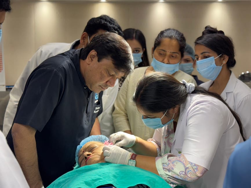
From an educational standpoint, Istanbul Dental Academy emphasizes clinical communication as a skill that can be practiced. Case-based discussions—combined with surgical simulation and supervised hands-on sessions—help clinicians learn how to present options without overpromising outcomes.
How related skills reduce sinus complications (even outside implant surgery)
While sinus perforations are strongly associated with implant dentistry and oral surgery, the preventive mindset is multidisciplinary. Conservative restorative dentistry can sometimes delay or reduce surgical needs by maintaining posterior tooth structure and occlusal stability. Likewise, predictable aesthetic-direct techniques can provide interim solutions when surgery must be staged.
For example, clinicians managing spacing or aesthetic concerns during surgical waiting periods may consider minimally invasive restorative strategies in suitable cases. The clinical benefits and limitations of such approaches are discussed in Advantages of Direct Composite Techniques for Closing Diastema. The key connection is not that diastema closure “treats” sinus issues, but that comprehensive care planning often requires coordinated surgical-restorative timing.
Training takeaways: translating knowledge into safer surgery
Knowing the theory of sinus perforation management is important, but translating that knowledge into predictable clinical performance requires repetition under guidance. In hands-on training, clinicians practice:
• CBCT-based risk assessment (sinus septa recognition, residual ridge measurement, pathology screening)
• Lateral window design and membrane elevation techniques with an emphasis on atraumatic instrumentation
• Complication algorithms for deciding when to repair and proceed versus stage and reassess
• Closure principles for extraction-related communications, including tissue management and prosthetic implications
Istanbul Dental Academy integrates these competencies into continuing dental education pathways that combine didactic sessions with practical workshops, helping clinicians build decision-making confidence—not just procedural familiarity.
Conclusion
Sinus perforations are not simply “bad luck”; they are often the predictable result of anatomical constraints, limited visualization, or incomplete planning. Prevention begins with CBCT-informed diagnosis and a disciplined surgical workflow. When perforations occur, early recognition and a structured, case-appropriate management plan can protect healing and long-term outcomes.
This content is for educational purposes and does not replace individualized diagnosis or treatment planning. For clinicians seeking to strengthen posterior maxillary surgery skills, complication management protocols, and digital planning competence, hands-on continuing education can be a practical next step.
Diğer Yazılar