BLOG
Case Selection for Porcelain Laminate Veneers: A Clinical Guide
Blog Tarihi: 14/06/2026

Why case selection matters more than “perfect veneers”
Porcelain laminate veneers can be among the most conservative and aesthetically powerful restorations in contemporary dentistry—when the right patient and the right tooth are chosen. In daily practice, many veneer failures are not caused by the ceramic itself, but by a mismatch between indications, periodontal and occlusal conditions, restorative limitations, and patient expectations.
For dental professionals and students, case selection is the step that transforms veneer therapy from “cosmetic dentistry” into a predictable restorative-prosthodontic procedure. This article outlines a clinically oriented approach to selecting cases for laminate veneers and highlights how structured diagnostics—supported by digital dentistry, dental photography, and interdisciplinary planning—reduces risk. This content is for educational purposes and is not a substitute for individualized clinical decision-making.
Start with the end in mind: patient goals and aesthetic objectives
Clarify the chief complaint and define success
Begin with a focused interview: What does the patient dislike—color, shape, asymmetry, spacing, wear, “small teeth,” or an uneven gingival line? Ask how long the concern has existed and what “ideal” looks like for them. Many patients arrive with references to popular smile makeovers; understanding these expectations helps guide material choice, tooth preparation strategy, and whether veneers are indicated at all.
When patients request an ultra-bright, high-value appearance, frame the conversation within a broader smile design workflow. You may find it useful to align the discussion with concepts from descriptive guidance on Hollywood Smile techniques and clinical workflow, especially when explaining how bleaching, alignment, restorative options, and soft-tissue considerations can be sequenced rather than rushed into veneers.
Set realistic boundaries early
Explain what veneers can and cannot predictably correct. Mild shape discrepancies, diastemas, enamel discolorations, and minor incisal wear often respond well. However, severe crowding, parafunction, unstable periodontium, and large existing restorations may shift the plan toward orthodontics, full-coverage restorations, or alternative adhesive strategies.
Core indications: when veneers are typically a strong choice
Laminate veneers commonly perform best when the clinician can bond predominantly to enamel and maintain additive or minimally invasive preparations. Typical indications include:
• Color and surface defects: fluorosis, enamel hypoplasia, localized discolorations where bleaching alone is insufficient.
• Shape and proportion issues: peg laterals, disproportionate width/length, minor asymmetries.
• Space management: small diastemas, black triangles (after careful periodontal evaluation), mild tooth rotation with additive contours.
• Incisal edge rehabilitation: limited chipping or wear when occlusion is favorable and protective strategies are in place.
From an educational perspective, these scenarios are excellent for teaching diagnostic waxing, mock-up transfer, reduction guides, and adhesive protocols—skills that Istanbul Dental Academy emphasizes in hands-on training environments where clinicians can rehearse the full workflow from photography to cementation.
Red flags and contraindications: when to pause or change the plan
Periodontal instability and gingival inflammation
Healthy soft tissues are not optional for veneers. Bleeding on probing, uncontrolled inflammation, and unstable gingival margins compromise impressions/scans, margin design, bonding field isolation, and long-term aesthetics (e.g., recession, “grey” margins, or exposure of cervical interfaces). Before finalizing veneer therapy, address periodontal disease and hygiene limitations.
Acute or necrotizing conditions require urgent periodontal evaluation rather than elective aesthetic dentistry. For a clinical refresher on high-risk inflammatory presentations, review Necrotizing Ulcerative Gingivitis (NUG): symptoms, causes, and clinical approach to contextualize why elective bonding procedures should be deferred until stability is achieved.
High caries risk and poor maintenance
Veneers do not “cure” caries activity. If the patient has active lesions, high sugar intake, xerostomia, or low compliance with home care and recall, the restoration margin becomes a vulnerable zone. Risk modification, preventive planning, and caries control should precede aesthetic rehabilitation.
Parafunction and unfavorable occlusion
Bruxism, clenching, and edge-to-edge bite relationships increase stress at the incisal edge and cervical areas, raising the likelihood of chipping, debonding, or ceramic fracture. Occlusal assessment should include guidance patterns, overjet/overbite, wear facets, mobility, and signs of muscle hyperactivity. In higher-risk cases, consider whether a different restorative design, material strategy, or protective occlusal splint plan is more appropriate.
Limited enamel and large existing restorations
Bond strength is generally more predictable to enamel than dentin. When teeth have extensive composite restorations, dentin exposure, or endodontic access with substantial structural loss, full-coverage restorations or alternative adhesive designs may provide better long-term performance. This is not absolute—experienced clinicians can manage complex cases—but it should be a conscious decision informed by risk-benefit discussion.
A structured diagnostic workflow for predictable case selection
1) Photography and baseline records
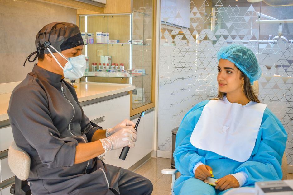
Standardized extraoral and intraoral photographs, shade mapping, and short video clips of speech and smile dynamics help translate patient expectations into measurable design goals. For clinicians, photography also provides objective documentation of gingival display, incisal edge position, midline, cant, and lip support—critical when planning conservative veneers.
2) Digital scans or high-quality impressions
Intraoral scans enable quick mock-up iterations, digital wax-ups, and communication with the laboratory. They can also highlight undercuts, path of insertion concerns, and interproximal morphology that influences emergence profile. In hands-on continuing education, practicing scan strategies and retraction techniques is often the missing link between theoretical smile design and chairside predictability.
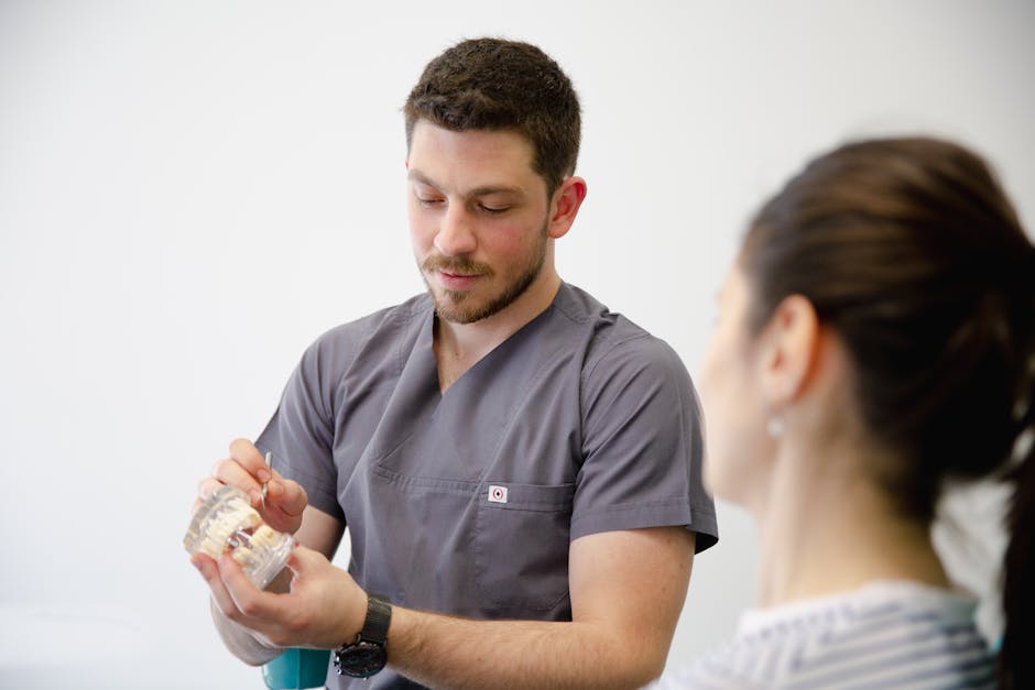
3) Diagnostic wax-up and mock-up (non-negotiable)
A diagnostic wax-up—analog or digital—lets you test tooth proportions, functional envelope, and occlusal contacts before irreversible preparation. Transfer the design intraorally with a mock-up and evaluate phonetics (“F/V” sounds for incisal edge length), lip posture, and patient comfort. This step frequently reveals when the patient’s aesthetic goal would require aggressive reduction and suggests alternative pathways such as orthodontics or bleaching first.
4) Periodontal evaluation and soft-tissue planning
Evaluate gingival levels, papilla fill, biotype, and smile line. If the patient has a high smile line, minor discrepancies in margins become more visible—making tissue management and margin placement even more critical. In selected cases, soft-tissue augmentation or periodontal plastic surgery can improve stability and aesthetics, particularly when recession risk or thin biotype is present.
For clinicians exploring biomaterials, clinical discussion of acellular dermal matrix use in dentistry can support a broader understanding of soft-tissue management concepts that may intersect with aesthetic restorative planning (case-dependent and requiring appropriate training and indications).
Preparation design: selecting cases where minimally invasive dentistry is realistic
Enamel preservation as a planning principle
Veneers are most predictable when the preparation stays in enamel across the majority of the bonding surface. Case selection should ask: Can we achieve the desired change additively, with minimal facial reduction? If the plan requires major changes in tooth axis, heavy masking of dark substrates, or significant incisal lengthening with heavy functional load, the preparation may become more invasive and the risk profile changes.
Margin location, isolation, and tissue response
Subgingival margins can complicate isolation and may challenge long-term periodontal health, especially in patients with inflammation history. Whenever possible, aim for supragingival or equigingival margins that respect the biologic width and allow excellent finishing and cleaning access. If shade changes require deeper margins, weigh the benefit carefully against periodontal and bonding challenges.
Material selection and bonding strategy: case selection continues here
Choosing lithium disilicate, feldspathic porcelain, or other ceramics depends on required translucency, thickness constraints, and strength needs. Thin, highly aesthetic feldspathic veneers may perform beautifully in low-stress anterior cases with abundant enamel and stable occlusion, while lithium disilicate may offer a more forgiving strength profile for moderate changes.
Even with ideal case selection, the clinical outcome hinges on adhesive detail. If your team is refining protocols, it is worth reviewing clinical tips on critical points in porcelain laminate cementation to reinforce how isolation, try-in paste selection, etching/silanization steps, and seating strategy impact long-term success.
Interdisciplinary considerations: when veneers are part of a bigger plan
Orthodontics and alignment before veneers
“Instant alignment” with veneers can be tempting, but excessive masking of rotations or crowding often demands aggressive reduction. When alignment is the root problem, orthodontic treatment may be the more conservative path, followed by selective bonding or minimal veneers for shape refinement.
Endodontics and structural integrity
Teeth with previous trauma, large restorations, or discoloration may require endodontic evaluation and a plan to manage substrate color (e.g., internal bleaching where appropriate) before committing to veneer thickness that could otherwise become overly invasive. A practical question for case selection: will the tooth remain structurally stable under functional load with a bonded partial-coverage ceramic?

Implant dentistry: managing mixed cases
Many smile makeover patients present with a combination of restorative needs: veneers on anterior teeth, crowns on heavily restored teeth, and implants to replace missing units. In such cases, the veneer plan should coordinate with implant prosthetics, emergence profiles, and midline/symmetry goals. When full-arch needs exist, it can be helpful to understand how implant planning choices affect the restorative envelope; see key differences between All-on-4 and All-on-6 for full-arch implant planning as a broader context for comprehensive treatment planning discussions (even if veneers are only one component).
A practical case-selection checklist (chairside-ready)
Patient factors
• Clear, realistic aesthetic goal; understands limitations and maintenance
• Acceptable oral hygiene and willingness for follow-up
• Caries risk assessed and controlled
Tooth factors
• Sufficient enamel for bonding across most surfaces
• Minimal existing restorations on facial surface (or planned strategy to manage them)
• Manageable discoloration without excessive thickness
Occlusal factors
• Functional guidance evaluated; no uncontrolled parafunction
• Adequate overjet/overbite for planned incisal lengthening
• Plan for protection if risk is moderate (case-dependent)
Periodontal factors
• Stable probing depths; minimal bleeding on probing
• Harmonized gingival margins relative to smile line
• Biotype and recession risk considered; soft-tissue plan when indicated
Workflow factors
• Diagnostic wax-up and intraoral mock-up completed
• Material and cementation protocol selected with isolation strategy
• Lab communication includes photos, shade mapping, and design parameters
Training note: why hands-on education changes outcomes
Case selection is a skill that improves dramatically with guided repetition: analyzing photos, evaluating occlusal schemes, preparing typodonts with reduction guides, and troubleshooting bonding steps under realistic isolation constraints. At Istanbul Dental Academy, veneer education is often integrated with smile design, digital workflows, and restorative-prosthodontic fundamentals—helping clinicians connect diagnostic decisions to practical chairside execution.
Conclusion
Predictable laminate veneer treatment begins long before the bur touches enamel. By prioritizing periodontal stability, enamel-preserving designs, occlusal risk assessment, and a mock-up-driven workflow, clinicians can select cases where veneers remain conservative, aesthetic, and maintainable. For dentists seeking to standardize their approach, structured documentation and hands-on training can bridge the gap between “good-looking cases” and repeatable clinical outcomes. This content is for educational purposes and does not replace individualized diagnosis or treatment planning.
Diğer Yazılar


