
BLOG
Myofascial Pain Syndrome in Dentistry: Evidence-Informed Care and Training
Blog Tarihi: 16/09/2026
Why myofascial pain syndrome matters in daily dental practice
Myofascial pain syndrome (MPS) is a common source of orofacial discomfort that can present as aching, pressure, fatigue, or “tooth-like” pain—often without clear odontogenic findings. For dental professionals, the challenge is not only symptom control, but also accurate differentiation from pulpal pathology, periodontal pain, neuropathic pain, and temporomandibular disorders (TMD). In busy clinics, MPS may appear in patients seeking restorative rehabilitation, smile design, implant treatment, or occlusal adjustments—especially when stress, sleep problems, or parafunctional habits (e.g., clenching) are in the background.
From an education perspective, MPS is a condition where clinical reasoning, hands-on palpation skills, and patient communication matter as much as technology. At Istanbul Dental Academy, we emphasise structured diagnostics and practical training because managing orofacial pain influences treatment planning across prosthodontics, periodontology, endodontics, oral surgery, and digital dentistry. This content is for educational purposes and does not replace individual diagnosis or treatment planning by a qualified clinician.
Clinical presentation: how MPS shows up in the dental chair
MPS is characterised by regional muscle pain associated with myofascial trigger points—hyperirritable spots within taut bands of skeletal muscle that can refer pain to distant sites. In dentistry, the muscles of mastication (masseter, temporalis, medial/lateral pterygoids) and cervical/shoulder muscles may be involved. Patients may report:
• Diffuse facial pain that worsens with chewing or prolonged speaking
• Morning jaw fatigue or stiffness, sometimes linked to bruxism
• Headache patterns (temporal or occipital), ear fullness, or neck discomfort
• Pain referral to posterior teeth, which can mimic endodontic symptoms
• Limited opening due to pain rather than true mechanical restriction
A key clinical pitfall is overtreating suspected tooth pain when the primary driver is muscular. Conversely, not every myofascial complaint is “just muscle.” A rigorous diagnostic pathway helps protect patients from unnecessary interventions and helps clinicians plan minimally invasive care.
Diagnosis: evidence-informed assessment and differential diagnosis
History taking: patterns that raise suspicion
In a dental history, ask about onset (sudden vs gradual), triggers (chewing, stress, posture), sleep quality, parafunction, recent dental procedures, and medication use. Explore psychosocial contributors without implying causation; stress and sleep disruption often coexist with pain but do not explain every case.
Clinical exam: palpation, function, and provocation
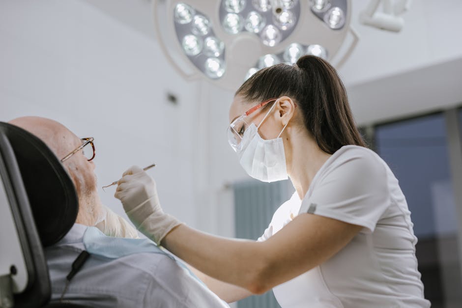
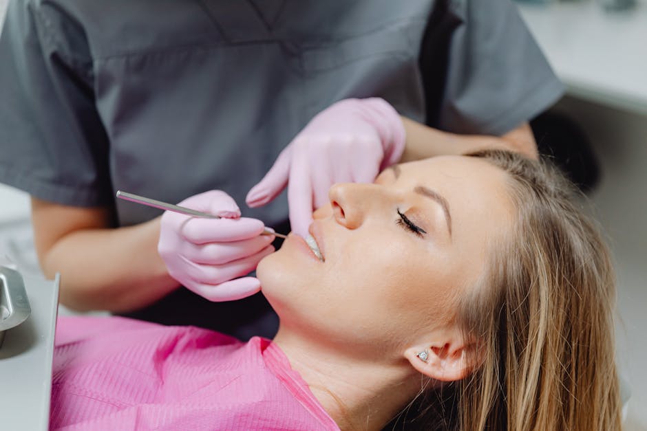
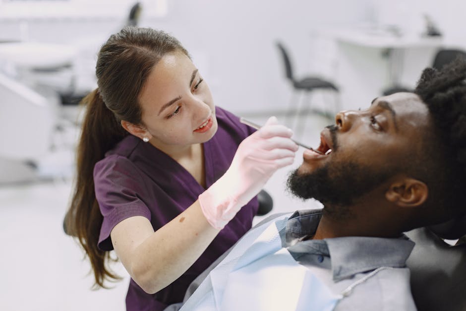
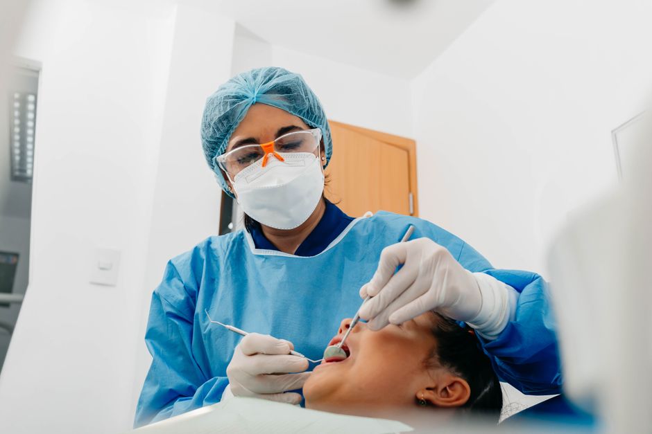
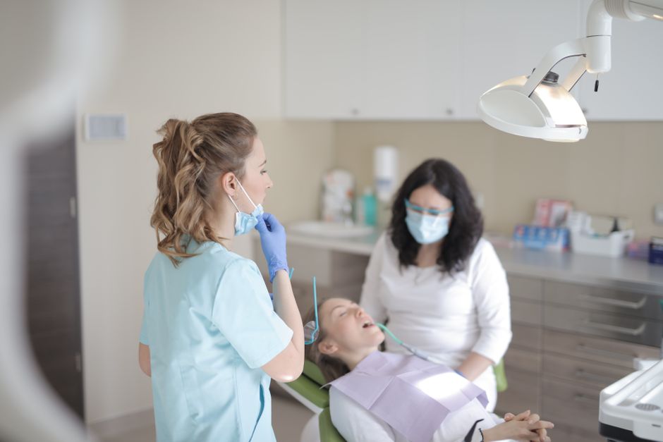
A structured exam typically includes observation (asymmetry, mandibular deviation), range of motion (active/passive opening), TMJ sounds, and muscle palpation with standardised pressure. Reproduction of the familiar pain during palpation and referred pain patterns can support an MPS hypothesis.
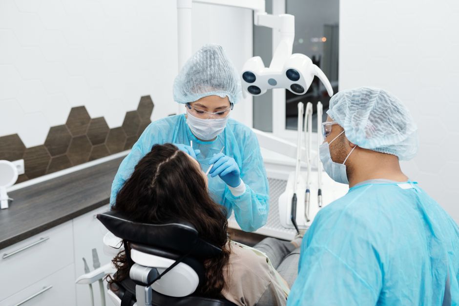
Because MPS often overlaps with TMD, a stepwise, evidence-informed diagnostic framework is essential. For a deeper clinical pathway focused on TMD patients, see Istanbul Dental Academy’s resource on a clinical diagnostic approach to TMD patients, which complements myofascial assessment and improves consistency in documentation and referrals.
Rule-outs that matter to dentists
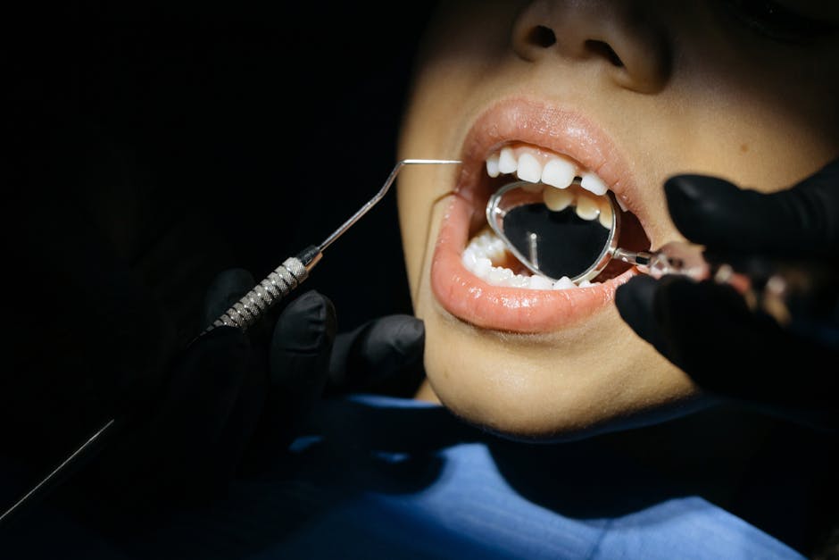
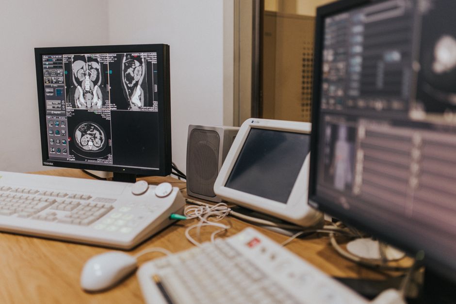
Responsible evaluation includes screening for odontogenic pain (pulpal testing, percussion, bite tests), periodontal pain (mobility, probing, occlusal trauma patterns), sinus-related symptoms, neuropathic pain clues, and red flags (rapid swelling, fever, progressive neurological deficits, unexplained weight loss). Imaging is not “routine” for MPS, but targeted imaging can be relevant when other conditions are suspected.
Contemporary management: a multimodal, conservative-first philosophy
Current care for MPS in the orofacial region tends to prioritise conservative, reversible strategies—often delivered through coordinated dental and medical collaboration. Treatment should be individualised and evidence-informed, with clear outcome measures (pain intensity, function, quality of life). The following options are commonly discussed in modern practice.
Patient education and self-management strategies
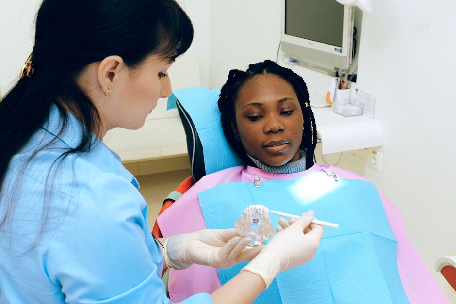
Education is not a “soft” intervention—it is frequently the foundation of sustainable improvement. Many patients benefit from guidance about jaw rest posture (lips together, teeth apart), limiting parafunctional behaviours, pacing chewy foods, and reducing prolonged mouth opening (including during dental appointments). Sleep hygiene and awareness of daytime clenching can be clinically relevant. Clinicians should frame advice as supportive and collaborative rather than prescriptive.
Physical therapy approaches and exercise
Physiotherapy may include gentle stretching, manual therapy, postural training, and home exercise programmes. For dentists, recognising when referral is appropriate—and how to communicate dental findings to physiotherapy colleagues—is a practical skill. In some cases, dry needling or trigger point techniques may be considered within the scope of local regulations and provider qualifications.
Occlusal appliances: careful indication and follow-up
Occlusal splints are commonly used when bruxism or muscle hyperactivity is suspected, yet they are not a universal solution. Appliance selection, design, and adjustment require careful assessment of symptoms, function, and patient compliance. A splint should be part of a broader plan, not a stand-alone “fix.” Clear follow-up intervals help ensure that an appliance is supportive rather than destabilising.
Pharmacologic options (supportive, not definitive)
Analgesics, anti-inflammatory medications, and muscle relaxants may be part of a broader management strategy under appropriate supervision. Dentists should follow local prescribing regulations and coordinate with the patient’s physician when relevant, especially for chronic pain, polypharmacy, or comorbidities. Medication is typically considered adjunctive—supporting comfort while functional rehabilitation and behavioural strategies address contributing factors.
Trigger point injections and botulinum toxin: selective use
In some settings, trigger point injections (e.g., with local anaesthetic) or botulinum toxin are used for selected patients, particularly when conservative approaches have not provided sufficient relief. Evidence and protocols vary by region, and clinicians should consider indications, contraindications, dosing strategies, and potential adverse effects (such as functional weakness). When used, these interventions are best integrated into a rehabilitation plan rather than replacing it.
Interdisciplinary links: periodontology, saliva, and pain patterns
MPS rarely exists in isolation. Periodontal inflammation, gingival recession sensitivity, and mucosal dryness can all influence how patients perceive pain and how they localise it. A patient with recession-related sensitivity may clench in response to discomfort, amplifying muscle pain, while chronic myofascial pain may heighten overall oral sensitivity.
When recession or dentin hypersensitivity is part of the complaint, it helps to evaluate the periodontal and behavioural context rather than focusing solely on symptoms. Istanbul Dental Academy’s overview of gum recession treatment options in modern dentistry can support a more comprehensive differential and patient conversation.
Salivary function also matters in orofacial comfort. Xerostomia can increase friction, alter oral tissue tolerance, and contribute to burning or soreness that patients may describe as “jaw pain.” If dryness, gland swelling, or medication-related salivary changes are suspected, a structured review is helpful. For clinical pointers, see a clinical guide to salivary glands and oral health.
Implications for restorative, prosthodontic, and aesthetic dentistry
In restorative dentistry and smile design, MPS can influence bite registration, jaw position consistency, and patient comfort during longer appointments. Muscular pain can alter mandibular posture, making centric relation records or occlusal evaluations less reliable. Patients may also attribute post-treatment discomfort to restorations even when the primary driver is muscular overload.
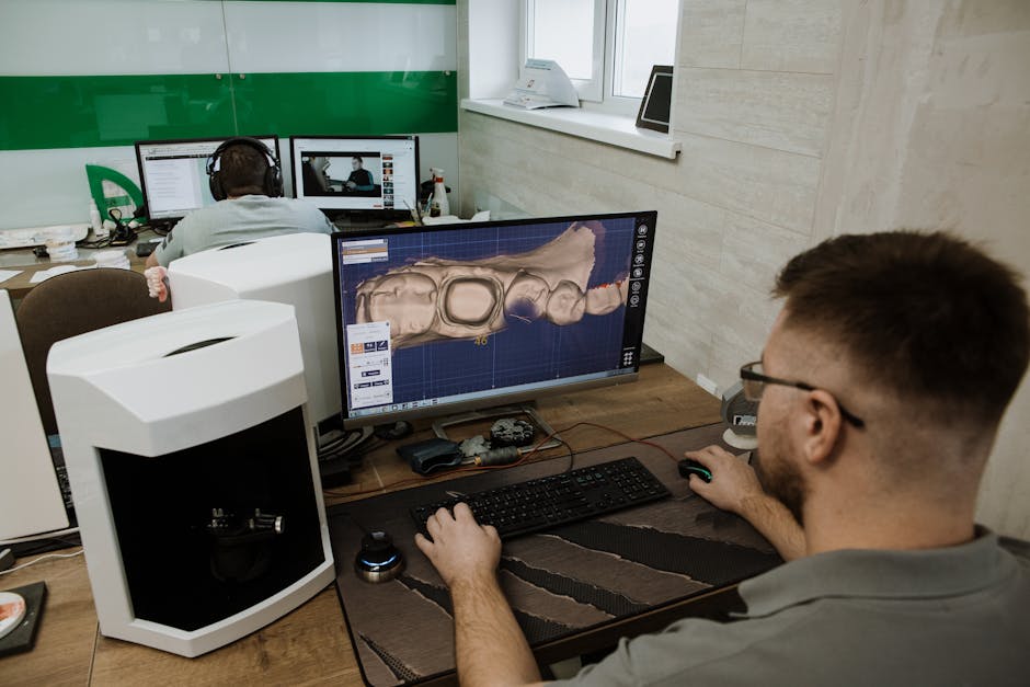
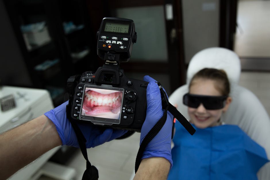
For porcelain laminate veneers and full-mouth rehabilitation, clinicians often benefit from phased planning: stabilise symptoms, confirm functional comfort, and proceed with minimally invasive steps when the patient’s muscle status is more stable. In digital workflows, this may include careful photographic documentation, jaw relation records, and staged provisionals to test function.
MPS considerations in implant dentistry and oral surgery
Patients seeking implants may present with chronic orofacial pain that predates tooth loss, or they may develop muscular guarding after extractions or surgery. While implant treatment is not a cause of MPS per se, prolonged mouth opening, stress-related clenching, and postoperative protective behaviours can exacerbate muscle tenderness.

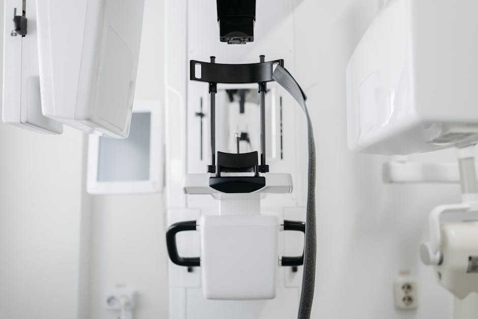
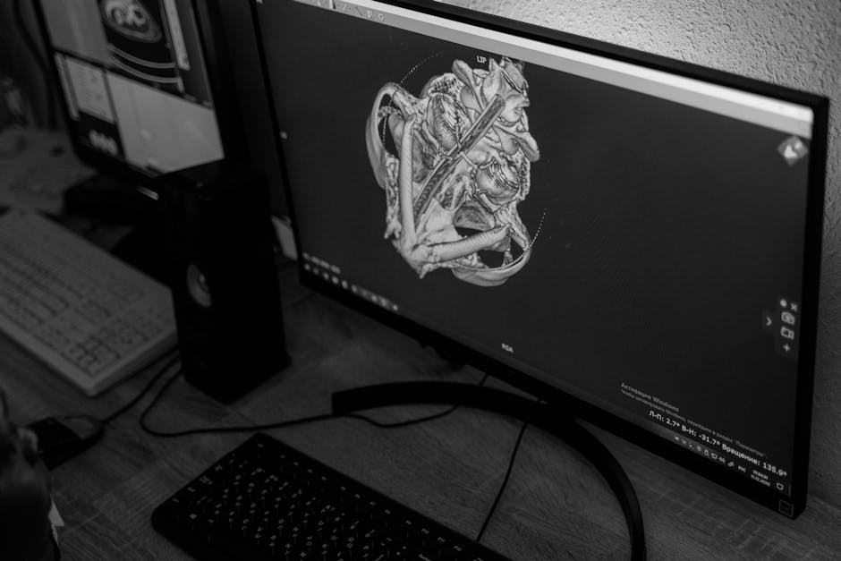
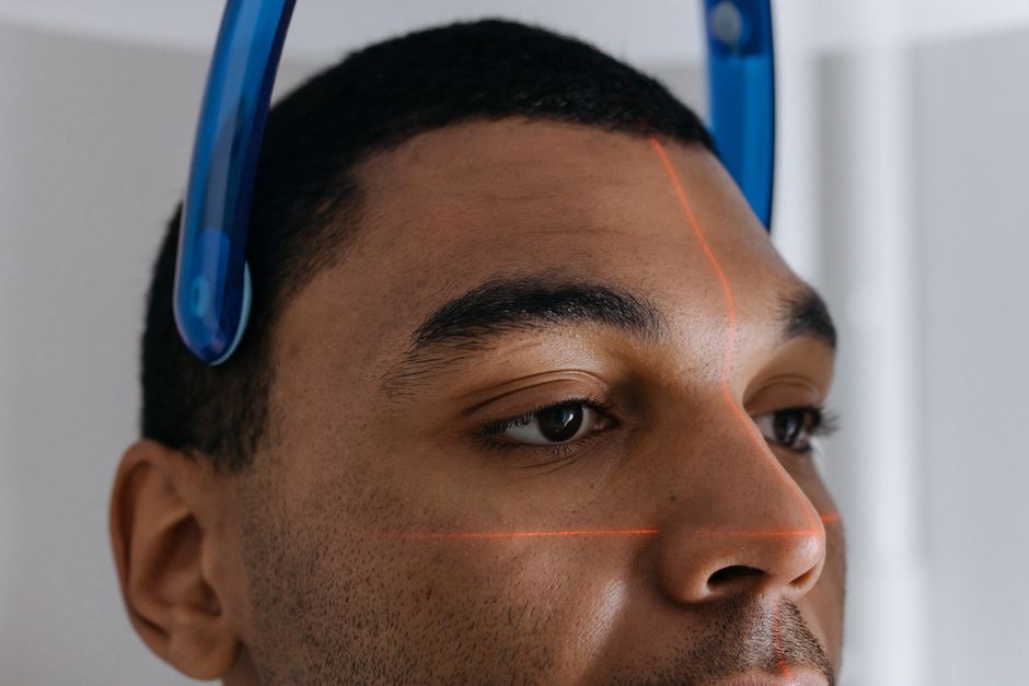
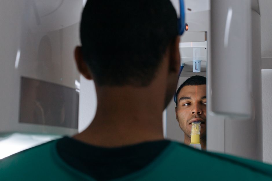
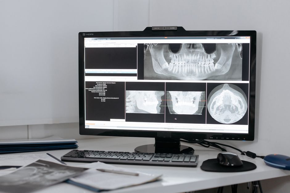
Risk management starts with planning and communication. In implant workflows, imaging and surgical strategy should be grounded in anatomy and patient-specific factors. Digital planning can reduce surprises that prolong chair time and strain the patient’s jaw. For a practical digital perspective, review why CBCT matters in dental implant planning, especially when anatomical limitations could complicate surgery and increase procedural time.
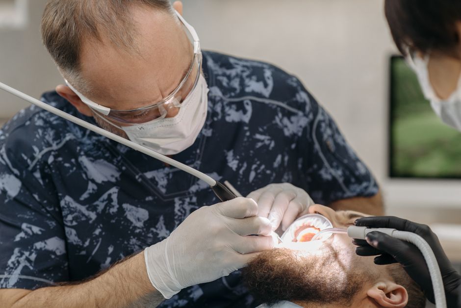
For early-career clinicians, another common issue is underestimating how technique and ergonomics influence patient comfort. Efficient, well-sequenced surgery can reduce unnecessary strain on the patient’s masticatory muscles. Istanbul Dental Academy discusses clinical pitfalls and prevention strategies in implant surgery mistakes beginners make—and how to prevent them, which indirectly supports better patient experience in individuals prone to muscle pain.
Digital dentistry and dental photography: documenting pain-related findings
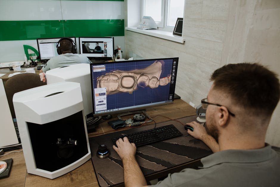
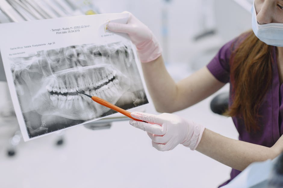
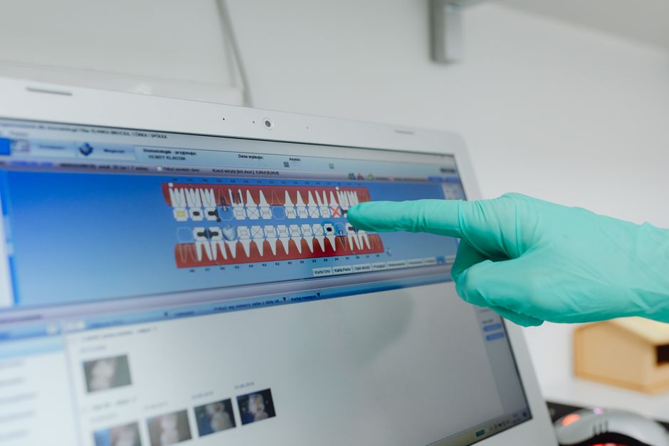
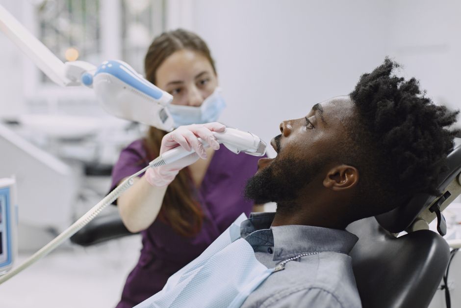
Although MPS is primarily a clinical diagnosis, documentation matters for continuity of care and interdisciplinary collaboration. Standardised extraoral and intraoral photographs can help track swelling, posture, asymmetry, and parafunctional wear patterns over time. Digital records also support patient education by making invisible factors (such as wear facets or tongue scalloping) more tangible.
In continuing education settings, combining photography protocols with structured exam forms helps dentists communicate findings clearly—especially when referring to physiotherapy, ENT, pain medicine, or oral and maxillofacial surgery colleagues.
How continuing dental education improves outcomes in orofacial pain cases
Managing MPS confidently requires more than reading: it depends on calibrated palpation, differential diagnosis, occlusal reasoning, and realistic patient communication. Hands-on training can refine these skills and reduce both over-treatment (e.g., unnecessary endodontics) and under-treatment (e.g., dismissing persistent pain without a plan).
At Istanbul Dental Academy in Istanbul, our continuing dental education philosophy is centred on clinic-ready learning: integrating diagnostics with practical workflows in restorative dentistry, prosthodontics, periodontology, and implantology. When dentists strengthen their examination consistency, digital planning, and documentation habits, they are better positioned to manage complex presentations where muscular pain overlaps with dental needs.
Key takeaways for dental professionals
• Think muscle when pain is diffuse or inconsistent with odontogenic tests. MPS can mimic toothache and complicate treatment planning.
• Use a structured TMD/MPS exam. Palpation, function tests, and careful history reduce misdiagnosis.
• Prefer conservative, reversible strategies first. Education, physiotherapy, and selective appliances are common components.
• Integrate periodontal and salivary context. Recession and xerostomia can influence symptom perception and habits.
• Optimise workflows for longer procedures. Digital planning and efficient surgery can reduce jaw strain in sensitive patients.
This content is for educational purposes only and does not constitute medical advice. Diagnosis and treatment should be personalised by qualified clinicians based on individual assessment.
Diğer Yazılar