BLOG
Why CBCT Matters in Dental Implant Planning: A Clinical, Digital Workflow Guide
Blog Tarihi: 25/06/2026
CBCT in Implant Planning: From “Where Can I Place It?” to “Where Should I Place It?”
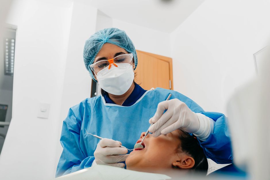
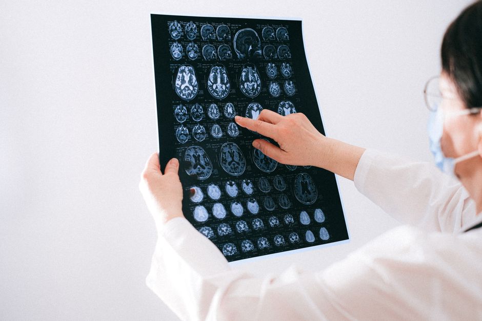
Dental implant dentistry increasingly depends on imaging that supports prosthetically driven decisions. Cone Beam Computed Tomography (CBCT) has become a key tool because it provides three-dimensional information that conventional two-dimensional radiographs cannot reliably offer in many cases. In daily practice, CBCT helps clinicians move beyond a purely bone-availability mindset toward a more comprehensive approach: respecting anatomical boundaries, planning restorative space, and anticipating soft-tissue and functional demands.
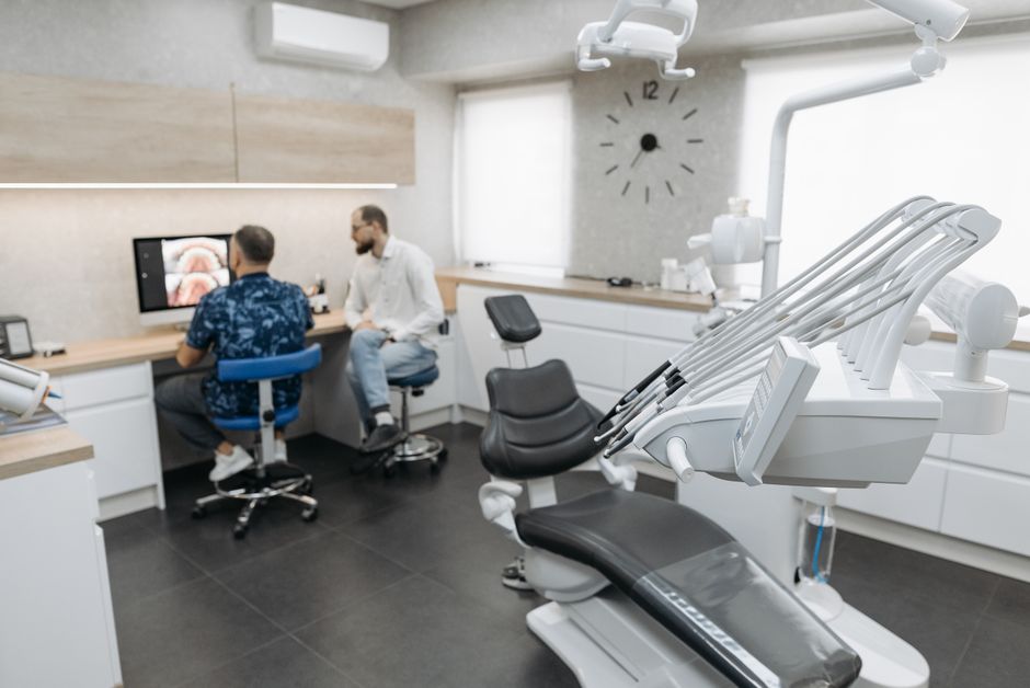
In Istanbul—where patient expectations for aesthetic, efficient, and digitally planned care are high—CBCT is often integrated into workflows that also include intraoral scanning, digital smile planning, and guided surgery. This article is for educational purposes and aims to clarify how CBCT supports clinical reasoning in implant planning, what it can and cannot do, and how training pathways can help clinicians use it responsibly.
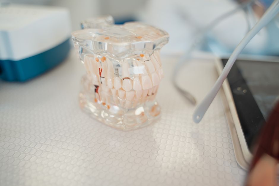
What CBCT Adds Compared with 2D Imaging
Panoramic and periapical radiographs remain essential in dentistry; however, they are inherently limited by magnification, distortion, and superimposition of anatomical structures. CBCT provides volumetric data that can be viewed in axial, coronal, sagittal, and cross-sectional slices—enabling more precise assessment of:
• Ridge morphology: buccolingual width, undercuts, concavities, and knife-edge ridges.
• Anatomical landmarks: inferior alveolar canal, mental foramen/anterior loop, maxillary sinus, nasal floor, incisive canal, and adjacent root proximity.
• Pathology and variations: cystic lesions, retained roots, sinus septa, and unexpected anatomical courses.
In prosthetically driven implant planning, these details directly influence implant diameter/length selection, angulation, need for augmentation, and restorative contour possibilities.
Key Clinical Questions CBCT Helps Answer
1) How much bone do we really have—3D?
Bone “height” on a panoramic image can be misleading if the ridge is narrow, concave, or irregular. CBCT cross-sections can reveal whether the clinician is facing a straightforward ridge or a morphology that may demand staged or simultaneous augmentation. This becomes particularly important in the anterior maxilla, where buccal plate thickness and concavity can affect both implant positioning and aesthetic outcomes.
2) Where are the risk zones?
Safe implant planning requires respect for anatomy. CBCT supports risk mapping, including the inferior alveolar canal and mental foramen in posterior mandible cases, sinus boundaries and septa in the maxilla, and the relationship to adjacent roots in narrow spaces. It can also help identify lingual undercuts that raise the stakes in the posterior mandible, where perforation risk may be underestimated in 2D views.
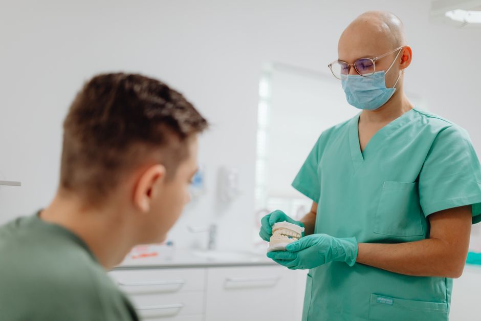
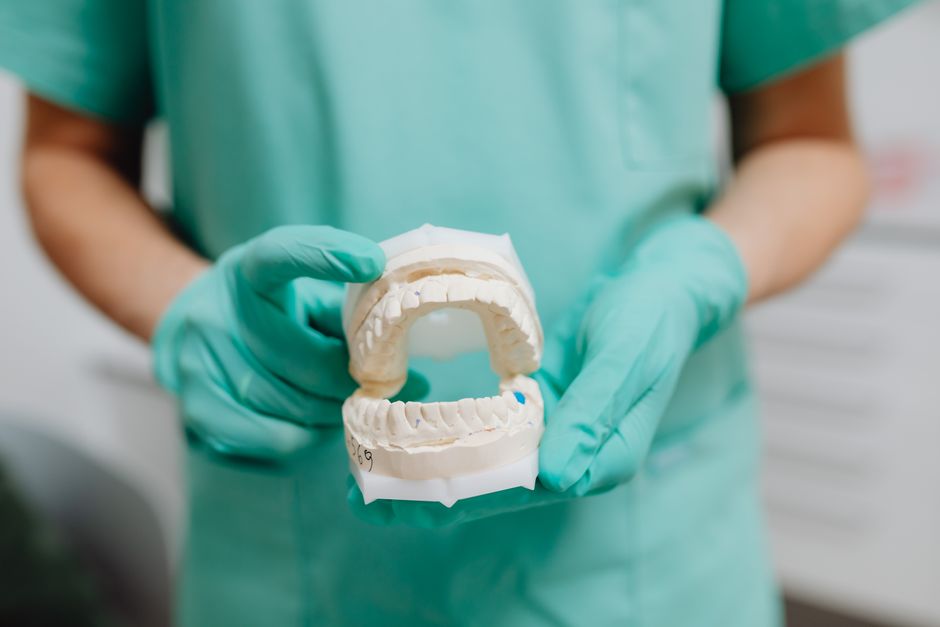
3) Can we plan the implant around the restoration?
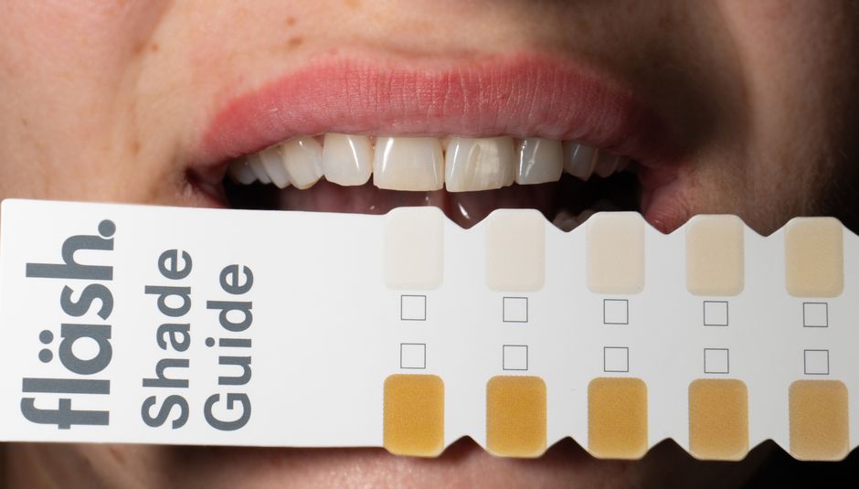
Implant placement is most predictable when planned backward from the final prosthesis. A digital workflow typically combines CBCT with intraoral scans or model scans so the clinician can visualize the bone and the planned crown simultaneously. This allows better decisions about emergence profile, screw access channel location, and restorative material thickness—especially relevant for high-demand cosmetic cases that may overlap with smile design workflows. Clinicians interested in how aesthetic planning integrates with restorative sequencing may also find value in reading What Is a Hollywood Smile? Techniques, Materials, and Clinical Workflow to understand how prosthetic goals can shape surgical decisions.
CBCT and Case Selection for Efficient Implant Protocols
Patients often ask about “same-day” solutions. While immediate implant placement and immediate loading can be appropriate in selected cases, CBCT plays an important role in evaluating whether anatomy and bone volume are compatible with such protocols. For instance, primary stability potential, socket morphology in immediate placement, and proximity to anatomical structures can be explored more thoroughly in a 3D dataset.
For an educational overview of how clinicians think through these indications and limitations, see Same-Day Dental Implants: Advantages, Limitations, and Case Selection. CBCT should be understood as one component of a broader diagnostic picture rather than a shortcut to aggressive timelines.
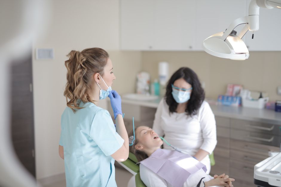
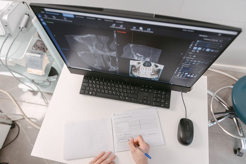
Planning with CBCT: A Practical, Digital Workflow
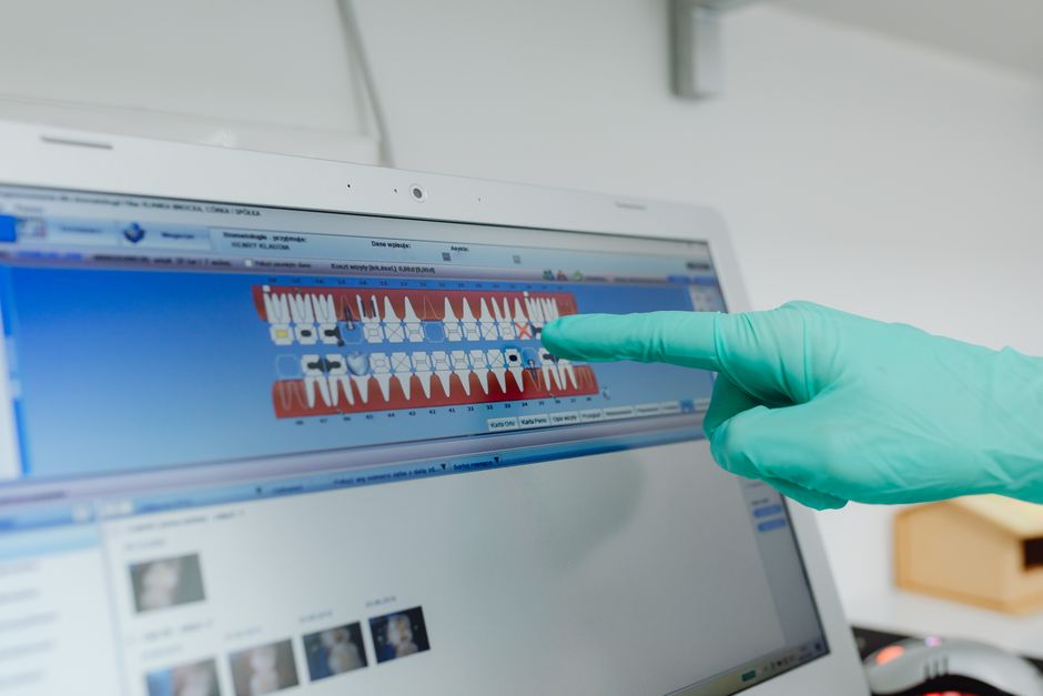
Step 1: Define the prosthetic endpoint
Before placing virtual implants, define crown position and occlusal scheme. This step may include digital smile design, diagnostic wax-ups, and photographic records. Aesthetic cases benefit from alignment between facial references, gingival margins, and restorative contours, which can be integrated into planning software when combined with surface scans.
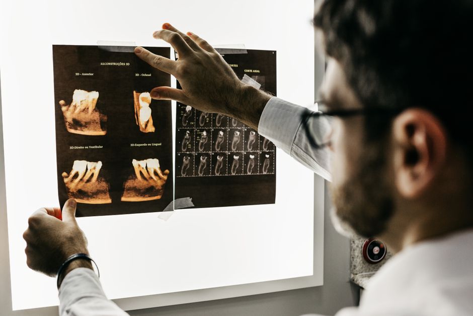
Step 2: Acquire a high-quality CBCT dataset
Accurate planning begins with accurate imaging. Field of view selection, voxel size, patient stabilization, and motion control all influence diagnostic value. The “best” settings are case-dependent; the guiding principle is to capture sufficient detail while following radiation safety principles and local regulations.
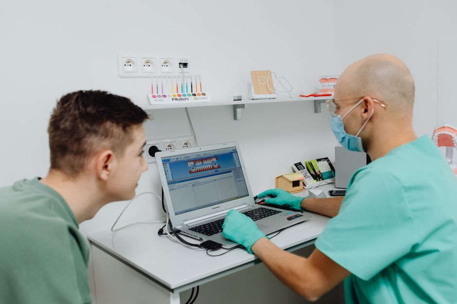
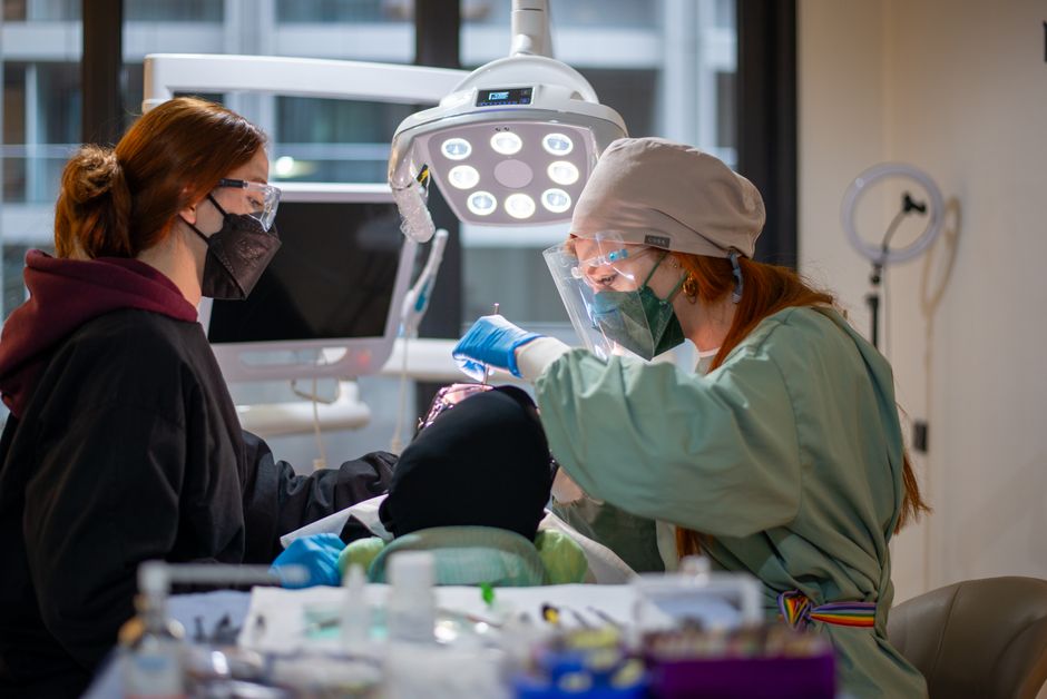
Step 3: Merge CBCT with intraoral scans (when indicated)
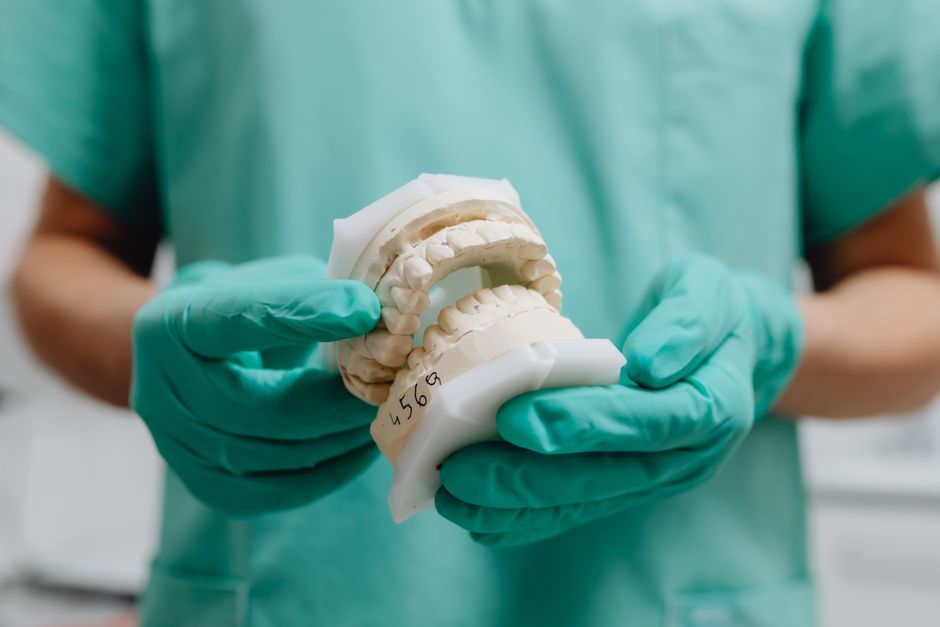
CBCT data provides bone and anatomy; intraoral scans provide teeth and soft-tissue surface detail. Their fusion enables more precise prosthetic-driven planning and is essential for most guided surgery workflows. In partially edentulous cases, stable tooth references usually make merging straightforward; in fully edentulous cases, scan protocols often require additional reference strategies.
Step 4: Virtual implant positioning and safety margins
Virtual planning typically involves selecting implant size, aligning the implant to the planned restoration, and confirming clearance from anatomical structures. Many clinicians plan with safety distances to key structures; however, the specific numbers and decisions should reflect the clinician’s training, the patient’s anatomy, and the limits of imaging resolution. This content is for educational purposes and does not replace clinical judgment or local guidelines.
Step 5: Decide on guided vs freehand surgery
Guided surgery can improve positional accuracy in many scenarios, particularly when combined with a prosthetically driven plan. Nonetheless, guides are not “autopilot”: they require correct seating, verification, and an understanding of tolerance and drill keys. CBCT-based guide design is often most beneficial in:
• narrow restorative corridors (e.g., anterior aesthetics),
• proximity to vital structures,
• immediate placement planning,
• full-arch cases where prosthetic alignment is critical.
CBCT Beyond Bone: Interdisciplinary Considerations
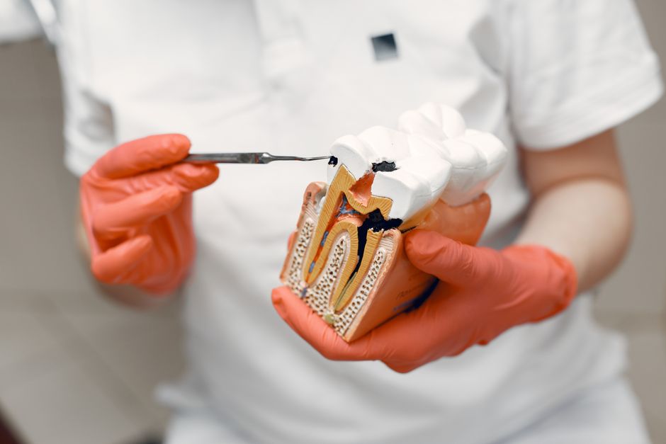
Although CBCT is widely associated with implant planning, its value often expands when cases involve periodontology, endodontics, or restorative dentistry. Examples include evaluating periodontal defects around potential abutment teeth, assessing endodontic pathology adjacent to an implant site, or diagnosing root fractures that could change the treatment plan. In complex cases, the imaging discussion becomes interdisciplinary: the “implant plan” may depend on periodontal stability, caries risk, occlusal loading, and maintenance capacity.
Why Oral Environment Matters: Saliva, Xerostomia, and Maintenance Planning
Implant success is not only about surgical placement; long-term maintenance and oral health behaviors matter. One often overlooked factor is salivary function. Saliva supports lubrication, buffering, antimicrobial activity, and mucosal comfort—elements that can influence plaque control and inflammation around teeth and implants alike.
If you want to revisit the systemic and local contributors to dryness and why it can matter clinically, read Dry Mouth (Xerostomia): Causes, Risks, and Clinical Implications. For a deeper foundation on diagnostic thinking and the broader oral implications, Salivary Glands and Oral Health: A Clinical Guide for Dental Professionals is a helpful companion piece.
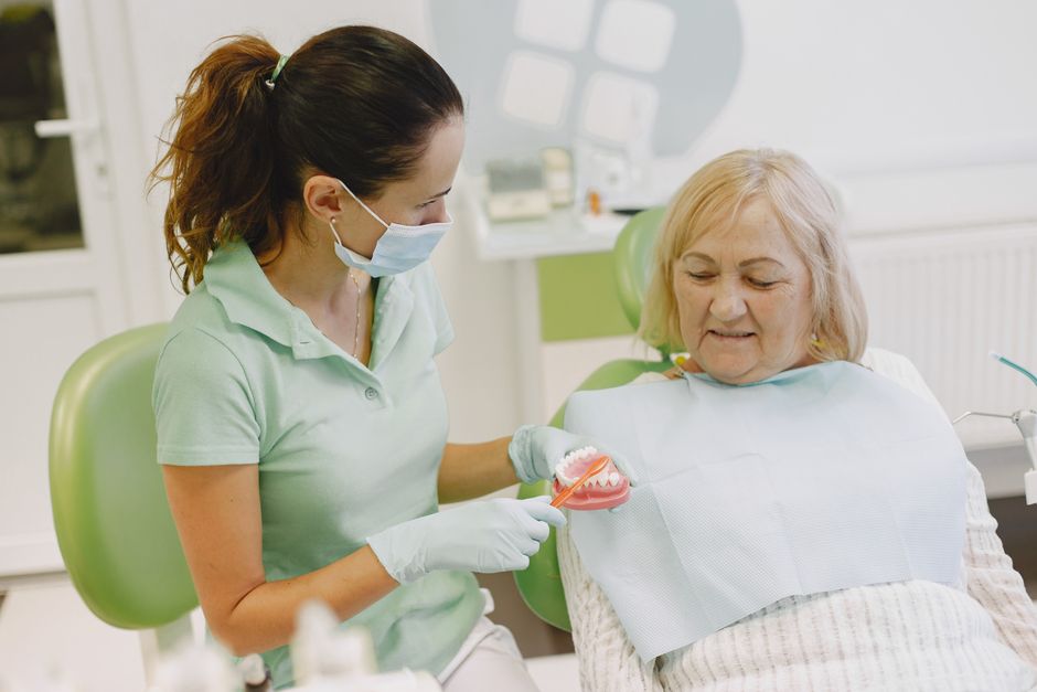
From a practical standpoint, clinicians often incorporate patient-specific maintenance planning into implant consultations: hygiene access, prosthesis cleansability, and risk-factor modification. When dryness is present, supportive strategies may be discussed as part of a comprehensive plan. For educational guidance on conservative approaches, see Natural Ways to Increase Saliva Flow: A Clinical Guide for Dental Professionals. These topics complement CBCT-based planning by addressing what happens after the scan—long-term tissue health and patient comfort.
Common Pitfalls: What CBCT Cannot Replace
CBCT strengthens diagnosis, but it does not eliminate uncertainty. Common pitfalls include:
Overreliance on “bone density” appearance: grayscale values in CBCT are not equivalent to medical CT Hounsfield Units in a standardized way. Clinical judgment about bone quality should integrate tactile feedback, surgical experience, and broader risk assessment.
Ignoring prosthetic space: placing an implant where bone exists but restorative space is inadequate can lead to compromised contours, hygiene challenges, and aesthetic limitations.
Underestimating soft-tissue impact: CBCT is not a substitute for soft-tissue evaluation. Biotype, keratinized tissue, and vestibular depth remain key clinical variables.
Skipping validation steps: in guided cases, failing to verify guide fit, stability, and sleeve/drill compatibility can negate the benefits of digital planning.
How Istanbul Dental Academy Approaches CBCT-Based Implant Education
For dental professionals and students, the major challenge is not accessing CBCT—but knowing how to interpret it systematically and translate findings into a safe, restorative-driven surgical plan. At Istanbul Dental Academy, implant education emphasizes structured reading of CBCT scans, digital planning principles, and clinically realistic decision-making, supported by hands-on training.
In a typical learning pathway, participants refine skills in:
• anatomical landmark identification and risk mapping,
• prosthetically driven implant positioning,
• guided surgery concepts and surgical guide evaluation,
• case selection (including immediate protocols when appropriate),
• documentation and communication using digital tools.
Hands-on components can help bridge the gap between “planning on the screen” and what occurs clinically—where visibility, access, and tissue behavior require adaptable skills.
Conclusion
CBCT has reshaped modern implant planning by enabling more confident 3D evaluation of bone and anatomy and by supporting digital, prosthetically driven workflows. When integrated thoughtfully—with clinical examination, restorative planning, and long-term maintenance considerations—CBCT can improve predictability and communication for both clinicians and patients.
This content is for educational purposes and is not a substitute for individualized diagnosis, treatment planning, or local regulatory guidance. For clinicians who want to strengthen their diagnostic and planning confidence, Istanbul Dental Academy’s continuing education and hands-on implant courses provide a structured way to turn CBCT data into practical, clinically relevant decisions.
Diğer Yazılar