BLOG
Critical Principles of Incision and Flap Design in Dental Surgery
Blog Tarihi: 27/06/2026
Why incision and flap design matters in everyday dentistry
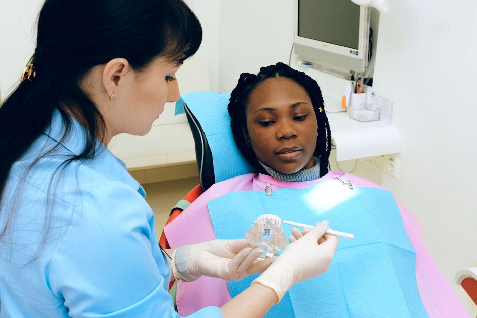
Incisions and flap design are often discussed as “surgical basics,” yet they frequently determine whether a procedure feels controlled and predictable—or stressful and compromised. In periodontal surgery, implant placement, ridge preservation, apical surgery, and even complex restorative workflows (e.g., subgingival margin management), the way soft tissues are approached affects access, visibility, haemostasis, postoperative comfort, and the long-term stability of the mucogingival complex.
From an educational perspective, incision and flap planning is where anatomy, biology, and technique intersect. Small decisions—where a releasing incision begins, how thick a flap is elevated, where tension is distributed—can influence wound stability, papilla preservation, and the risk of recession or scarring. For clinicians developing surgical confidence, these principles are best learned in structured continuing education with step-by-step planning, mentored repetition, and hands-on simulation—an approach Istanbul Dental Academy emphasizes across its surgical and restorative training pathways.
This content is for educational purposes and does not replace clinical judgment, diagnosis, or patient-specific treatment planning.
Biological foundations: blood supply, tissue thickness, and wound stability
Preserving vascularity is not optional
Soft tissues heal predictably when vascular supply is respected. Incision placement should avoid creating long, narrow tissue “tags” and should aim to maintain broad-based flaps with adequate perfusion. When flaps are undermined, thinned excessively, or stretched under tension, perfusion can be compromised—leading to delayed healing, dehiscence, or recession.
Understanding the soft tissue biotype and the patient’s baseline periodontal phenotype is equally important. Thinner tissue types tend to show more recession after trauma or tension. When clinicians see a case with pre-existing mucogingival challenges, it can be helpful to revisit the broader clinical picture of recession risk factors and management strategies, such as those discussed in Gum Recession: Causes, Symptoms, and Evidence-Based Management.
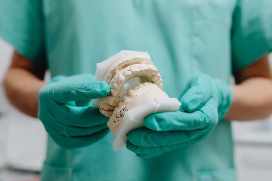
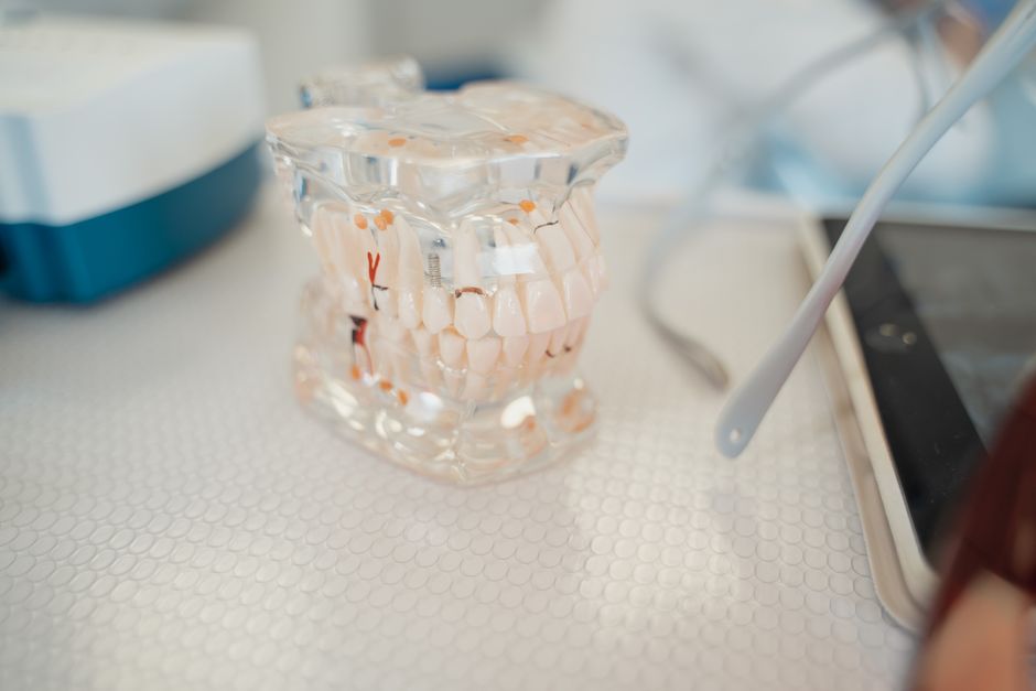
Thickness and flap type: full-thickness vs. split-thickness
Full-thickness (mucoperiosteal) flaps offer predictable access to bone and are commonly used in implant and oral surgery. Split-thickness flaps may be selected when periosteum preservation is desired for specific mucogingival procedures or to enable coronal advancement. The “correct” choice depends on the objective: access, augmentation, primary closure, or papilla preservation.
A useful teaching point in hands-on courses is to plan the flap based on the closure goal before the first incision is made. In other words: design the flap backward from the desired, tension-free closure line rather than improvising after reflection.
Incision planning: practical principles for predictable access
Start with the endpoint: what do you need to see?
Before selecting an incision, define the minimum access required for the planned steps: osteotomy preparation, debridement, graft placement, membrane adaptation, root-end preparation, or suturing. Overly conservative access can increase iatrogenic trauma due to retraction and instrument leverage. Conversely, overextending incisions can increase postoperative morbidity and scar risk.
Common incision patterns in clinical use
While nomenclature varies, clinicians typically choose between sulcular (intrasulcular), crestal, papilla-sparing, and paramarginal incision designs, often combined with one or two vertical releasing incisions. Each approach has trade-offs:
Sulcular incisions may offer excellent access and ease of repositioning but can threaten papilla stability if closure is under tension.
Crestal incisions are common in implant surgery; however, keratinized tissue management must be considered, especially in esthetic zones.
Paramarginal incisions can preserve marginal tissues but may limit access and complicate flap advancement in certain anatomies.
Releasing incisions: minimize tension and protect papillae
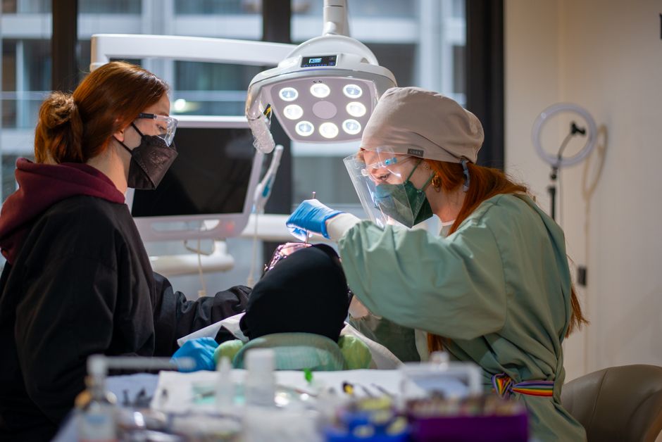
Vertical releases can improve mobility, but they also introduce additional wound edges and potential scarring. In many hands-on surgical trainings, clinicians learn to ask two questions before adding a release: (1) Is flap mobility truly insufficient without it? (2) Can a different advancement method (e.g., periosteal scoring) achieve tension-free closure with fewer incision lines?
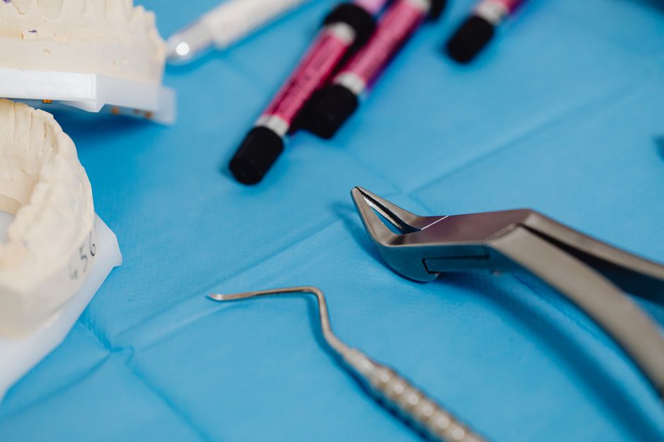
When releases are used, designs that avoid crossing prominent frena and that protect papillary blood supply are generally preferred. Incisions placed over bony eminences can be more prone to dehiscence. Precise execution matters: sharp instruments, a single confident stroke, and atraumatic handling reduce tissue crush injury.
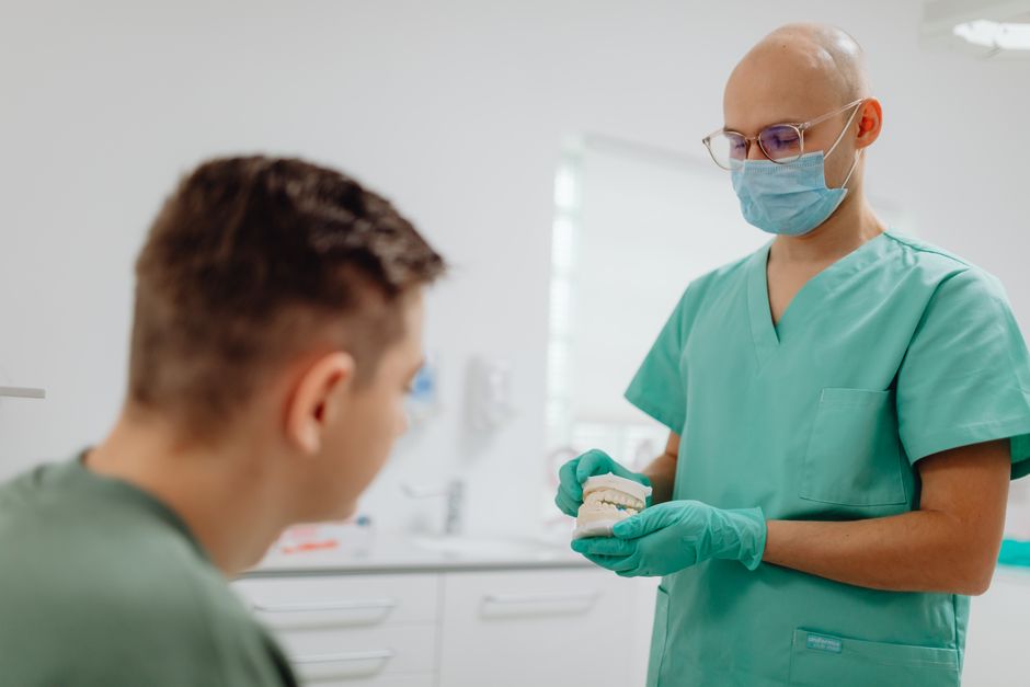
Flap design in implant dentistry: access, accuracy, and closure
Balancing visibility with soft-tissue stability
In implant dentistry, flap design impacts the surgeon’s ability to position implants prosthetically and to manage hard/soft tissue contours for long-term maintenance. Flapless approaches can reduce morbidity in selected cases, but they demand accurate preoperative planning and sufficient keratinized tissue. When the anatomy, ridge form, or restorative plan requires visualization and augmentation, a well-designed flap remains the workhorse.
Clinical discussions around accelerated protocols often raise questions about how much surgery can be done “quickly” while still respecting biology. If you are exploring time-efficient implant workflows, consider the case selection and surgical realities addressed in Is One-Day Dental Implant Treatment Really Possible?, where flap management and predictability are part of the broader decision-making process.
Immediate implant placement: extraction site realities
When placing an implant immediately after extraction, flap design can influence socket visualization, debridement, and the ability to achieve primary closure (when indicated). Some protocols aim for minimal flap elevation to preserve blood supply to the buccal plate and papillae, particularly in the anterior zone. Other scenarios—such as thin buccal plates, infection management, or planned grafting—may call for controlled flap reflection to confirm bony walls and graft containment.
For clinicians refining these decisions, Same-Day Tooth Extraction and Immediate Implant Placement: A Clinical Guide provides an educational overview that pairs well with hands-on training, where flap design is practiced alongside extraction biomechanics and suturing strategies.
Periodontal and restorative interfaces: papilla preservation and margin control
Why papilla management is an esthetic priority
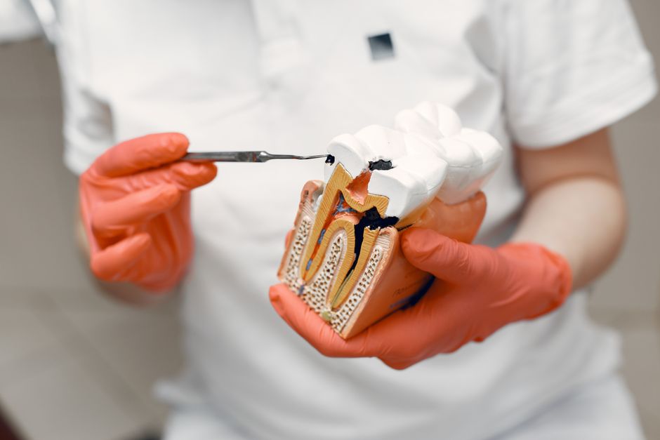
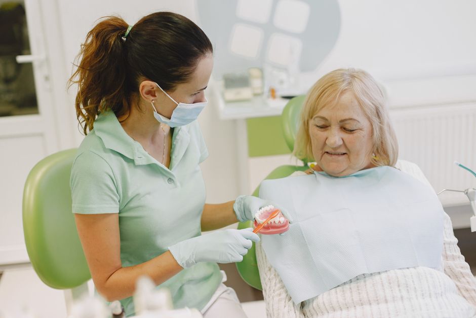
In the esthetic zone, the interdental papilla and marginal gingiva influence smile harmony as much as tooth form and color. Incisions that split or traumatize papillae can compromise black triangle management and soft tissue symmetry. In periodontal plastic surgery and crown lengthening, flap design is often the difference between a stable gingival margin and a drifting, unpredictable outcome.
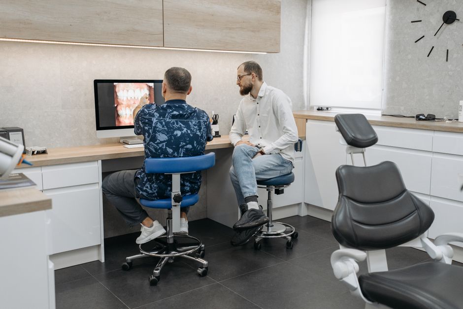
For clinicians integrating surgical concepts into restorative workflows (such as veneer preparations, smile design, and margin placement), the message is consistent: plan incisions around future tissue position and restorative emergence profiles. Many advanced programs at Istanbul Dental Academy connect these disciplines, using clinical photography and digital planning to map incision lines to the final restorative outcome.
Moisture control and oral environment considerations
Soft tissue behavior is also influenced by the oral environment, including salivary flow and mucosal health. While saliva is essential for healing and lubrication, altered salivary function can affect patient comfort, plaque control, and tissue response during postoperative phases. For a broader clinical refresher, see Salivary Glands and Oral Health: A Clinical Guide for Dental Professionals, which contextualizes salivary considerations relevant to surgical and restorative care.
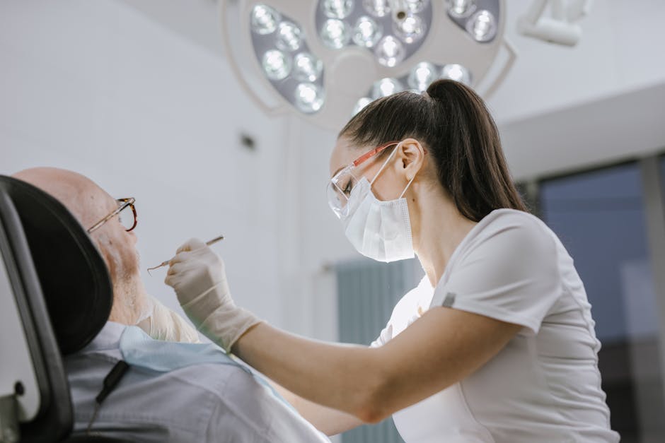
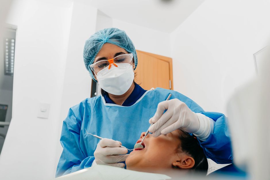
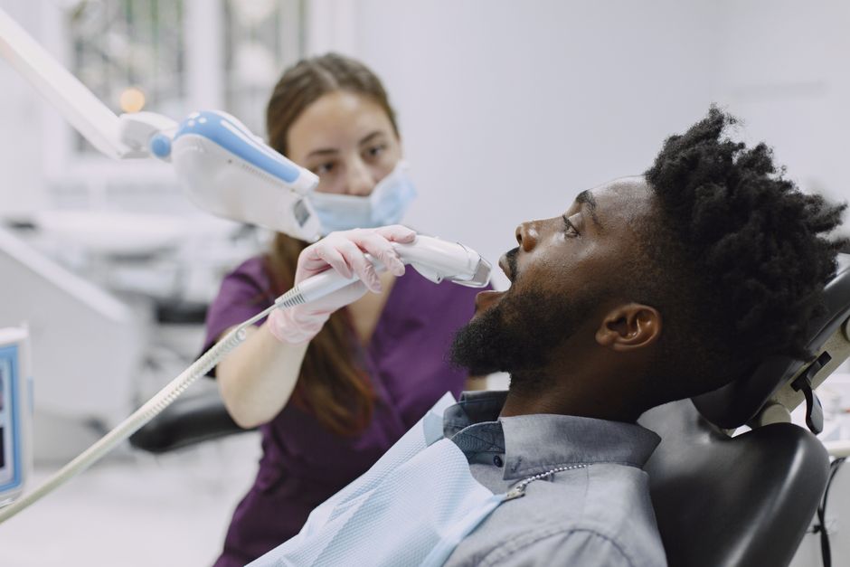
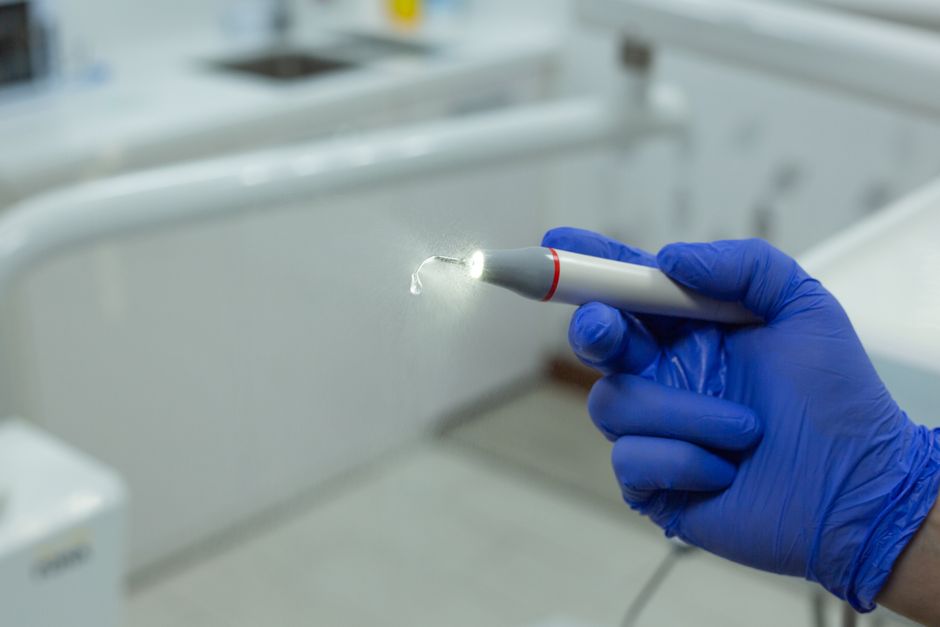
Microsurgical mindset: visibility, magnification, and precision
How magnification changes incision quality
Incision and flap design are not only conceptual—they are technical. Magnification can improve precision of incision placement, reduce unintended tissue tearing, and support more controlled suturing. Although microscopes are commonly associated with endodontics, the underlying benefit—enhanced visualization for fine motor control—translates to many surgical tasks.
If you are interested in how magnification supports clinical ergonomics and precision, Advantages of Using a Dental Microscope in Endodontics is a useful educational resource. Many clinicians find that adopting a “microsurgical mindset” improves their flap handling and wound-edge approximation even outside endodontic procedures.
Common pitfalls and how to avoid them
1) Designing a flap that cannot close without tension
One of the most frequent problems is planning for access but not for closure. If closure requires stretching tissue, the risk of dehiscence increases, particularly over grafted sites or thin buccal plates. In training, this is where suturing practice and periosteal release techniques become essential—not as “extras,” but as core skills that complete the surgical plan.
2) Over-retraction and tissue crush injury
Even a perfectly placed incision can be compromised by aggressive retraction. Excessive pressure reduces perfusion and can tear flap margins. Using appropriately sized retractors, stabilizing the hand, and limiting retraction duration are small changes with large outcomes.
3) Ignoring anatomical landmarks
Flap design should consider frena, muscle pull, vestibular depth, and neurovascular structures. A releasing incision placed where mobile mucosa dominates can increase postoperative discomfort and wound movement. This is a key reason why anatomic review and supervised practice are central to continuing dental education in surgery.
4) Underestimating biotype and recession risk
Thin phenotypes and existing recession demand more conservative tissue handling and thoughtful incision placement. Clinicians should anticipate where the margin might move after healing and avoid creating conditions that accelerate recession. When in doubt, a multidisciplinary conversation—periodontology, prosthodontics, and surgery—often leads to more stable planning.
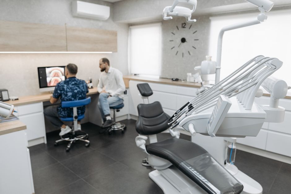
How Istanbul Dental Academy supports skill transfer: from theory to hands-on competence
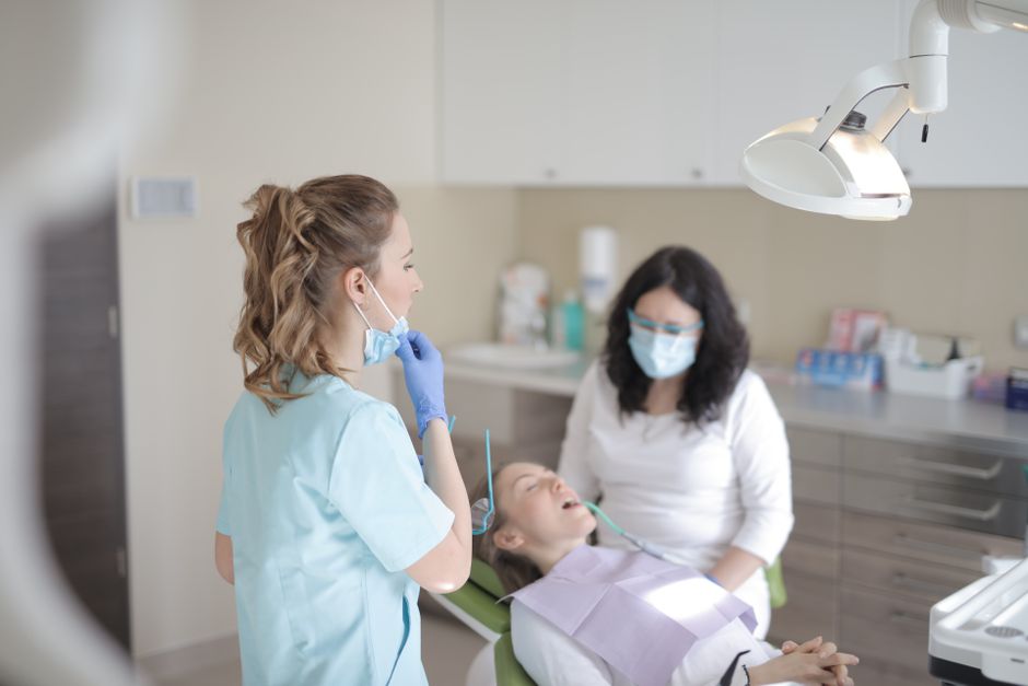
Reading principles is valuable, but surgical skill depends on repetition: instrument control, incision angulation, flap elevation planes, tension management, and suturing patterns. Istanbul Dental Academy’s educational philosophy prioritizes practical learning—hands-on dental courses, live demonstrations, guided exercises, and case-based discussions that connect incision/flap planning to real-world outcomes in implant dentistry, periodontal surgery, and restorative-driven treatment planning.
In a structured course environment, participants can practice how different flap designs behave during reflection and closure, how to adapt designs for immediate implant placement versus staged approaches, and how to document soft tissue changes through clinical photography and follow-up. The goal is not a one-size-fits-all “recipe,” but a repeatable planning framework clinicians can apply to their own cases.
Take-home planning checklist (educational)
1) Define the surgical objective: access, debridement, augmentation, or closure priority?
2) Evaluate phenotype and keratinized tissue: anticipate recession and margin shift risk.
3) Map incision lines to closure: plan tension-free approximation before cutting.
4) Protect vascularity: broad-based flaps, minimal crushing, cautious releases.
5) Choose the right visualization strategy: lighting, loupes, and magnification improve precision.
When these fundamentals become routine, incision and flap design transforms from a “basic step” into a controlled strategy that supports predictable healing across implant, periodontal, and oral surgery workflows.
Diğer Yazılar