BLOG
Minimal-Invasive Aesthetic Dentistry with Porcelain Laminate Veneers
Blog Tarihi: 25/06/2026
Why laminate veneers remain central to minimal-invasive aesthetic dentistry
In contemporary restorative dentistry, “minimal invasive” is more than a trend—it is a philosophy that prioritizes biological respect, enamel preservation, and long-term maintainability. Porcelain laminate veneers (often simply called “lamina veneers”) fit naturally within this approach because they can deliver high-impact aesthetic change with limited tooth reduction when the case is selected and executed properly.
For dental professionals, veneers also represent an intersection of disciplines: adhesive dentistry, occlusion, periodontology, prosthodontics, and digital planning. That interprofessional nature is why veneer success is strongly tied to systematic workflows—records, mock-ups, tooth preparation design, bonding strategy, and post-operative maintenance—rather than a “single-step” restorative mindset.
This content is for educational purposes and is not a substitute for individual clinical judgment, diagnosis, or treatment planning.
Case selection: indications, contraindications, and risk assessment
Common indications
Porcelain laminate veneers are frequently considered for:
• Shape and proportion corrections (peg laterals, incisal wear, uneven edges)
• Color modification (when whitening alone is insufficient or unstable)
• Mild-to-moderate alignment discrepancies (selected cases where orthodontics is declined or not feasible)
• Diastema closure and smile line optimization
• Additive rehabilitation of worn anterior teeth when enamel can be preserved
Key contraindications and “yellow flags”
Minimally invasive veneers become more complex when risk factors are present. Examples include parafunction, unstable occlusion, limited enamel for bonding, high caries risk, and uncontrolled periodontal inflammation. Soft tissue stability is particularly important: the best ceramic margins will not “save” an unstable gingival environment.
If recession or thin biotype is observed, veneer planning should be coordinated with periodontal assessment. A focused review of etiology and management considerations can be found in our educational article on gum recession and evidence-based management, which highlights why tissue phenotype and inflammation control matter for long-term aesthetics.
Diagnosis and records: the foundation of predictable aesthetics
Photography and communication
High-quality dental photography is not “extra”—it is the language of aesthetic diagnosis and lab communication. A consistent photo set (full face, smile, retracted views, occlusal mirrors, shade tabs) supports analysis of midline, incisal plane, buccal corridors, tooth texture, and color mapping. For clinicians building aesthetic workflows, learning controlled lighting and standardized angles significantly reduces remakes and chairside adjustments.
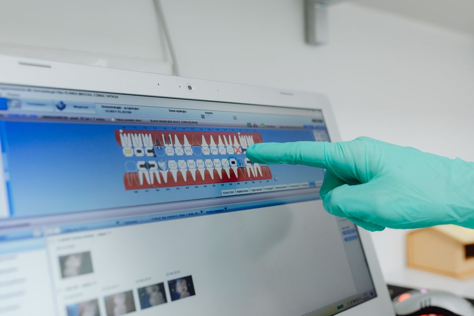
Digital smile design and mock-ups
Digital planning can be helpful in visualizing tooth proportions and gingival symmetry, but it should not replace functional evaluation. A diagnostic wax-up and intraoral mock-up remain valuable because they test phonetics (“F/V” sounds), incisal edge position, and patient acceptance—before enamel is touched.
Digital workflows also create a natural bridge between aesthetic dentistry and implant prosthodontics. The same planning logic—start with the final prosthesis, then design the supporting structures—appears in full-arch implant cases. Clinicians interested in structured digital protocols may also review digital planning for full-arch implant cases to see how data integration (CBCT, scans, and prosthetic planning) supports predictability in more complex rehabilitations.
Preparation design: preserving enamel while respecting ceramic principles
Additive vs minimally reductive preparations
A “no-prep” veneer is not automatically conservative if it creates over-contour, compromises cleansability, or produces an unnatural emergence profile. The true goal is enamel preservation while maintaining appropriate ceramic thickness and contours. Many successful cases use a minimally reductive approach (often 0.3–0.7 mm facial reduction depending on material, shade change needs, and tooth position), guided by a mock-up reduction index.
Finish line and margin placement
Margin placement is case-dependent: equi-gingival margins may be chosen for aesthetics, while supragingival margins can improve hygiene and tissue health when feasible. Subgingival margins may be considered selectively, but they increase technique sensitivity and soft tissue risk. Good soft tissue management is therefore a prerequisite, not an afterthought—especially in patients with inflammation, recession tendencies, or high aesthetic demands.
Incisal design and functional considerations
Incisal overlap (wrap) can enhance strength and aesthetics in some cases, but it must be coordinated with guidance patterns and parafunction risk. Anteriors that carry heavy lateral loads may require occlusal management, protective appliances, or alternative restorative strategies. Veneers are not a universal solution; they work best when the occlusal environment is stable or can be stabilized.
Material selection: why porcelain is often preferred
Porcelain laminate veneers are widely used due to optical stability, surface gloss retention, and wear compatibility when finished correctly. Lithium disilicate is common for many veneer cases because it balances translucency and strength while enabling adhesive bonding. Feldspathic ceramics may offer excellent aesthetics in thin sections but can be more technique-sensitive and may require careful case selection.
From an educational standpoint, clinicians benefit from learning how prep depth, shade goals, and underlying tooth color influence ceramic selection and cement shade strategy. In hands-on settings, practicing try-in protocols, cement selection, and cleanup timing can be as important as learning preparation geometry.
Adhesive bonding: where minimal invasion either succeeds—or fails
Enamel-driven bonding strategy
One of the strongest arguments for minimal reduction is the reliability of enamel bonding. Preserving enamel increases bond durability and reduces the risk of marginal staining and debonding over time. When dentin is exposed, bonding can still be successful, but the technique becomes more sensitive to isolation, adhesive choice, and operator consistency.
Isolation and cementation workflow
Rubber dam isolation is often considered best practice for adhesive cementation when possible. If rubber dam placement is limited by the case, alternative isolation strategies must be meticulous. Cementation includes proper ceramic pretreatment (etching for glass ceramics, silanization), tooth surface conditioning, controlled seating pressure, and stepwise tack-curing with careful cleanup to protect gingival tissues.
These are also the steps where many clinical complications originate: incomplete seating from excess cement viscosity, marginal gaps from rushed cleanup, or soft tissue inflammation from residual resin. In continuing dental education, repeated supervised cementation practice is one of the fastest ways to improve predictability and efficiency.
Veneers are not always the answer: conservative alternatives to consider
Minimal invasive dentistry prioritizes choosing the simplest restoration that fulfills the patient’s functional and aesthetic needs. In some posterior or cusp-related defects, partial coverage restorations may be more appropriate than extending indications beyond veneers.
For example, when cuspal coverage and occlusal risk are central concerns, an onlay or overlay may provide a more biomechanically sound solution. A structured comparison can be found in clinical decision-making for onlay vs overlay restorations, which can help clinicians align preparation design with long-term load management.
Integrating veneer planning with periodontal and endodontic considerations
Periodontal health and emergence profile
Aesthetic dentistry is inseparable from periodontal stability. Over-contoured veneers can trap plaque and provoke inflammation, while overly aggressive subgingival margins can destabilize tissue levels. Evaluating tissue phenotype, keratinized tissue, papilla height, and smile line helps determine whether restorative-only treatment is realistic or whether periodontal co-management is required.
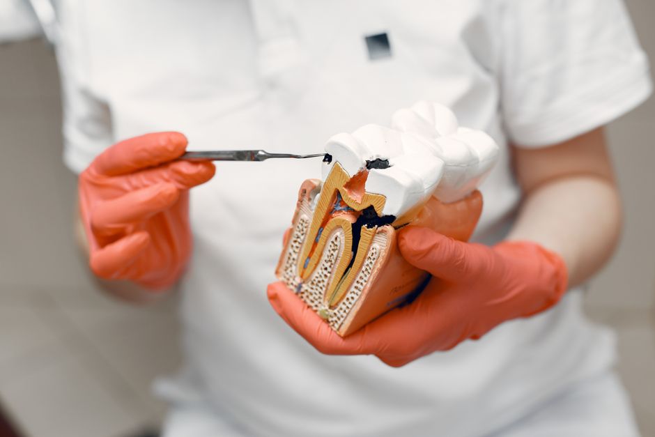
Teeth with previous endodontic treatment
Endodontically treated anterior teeth can be candidates for veneers in selected cases, but discoloration, remaining tooth structure, and risk of fracture should be considered. Some situations may require internal bleaching, core optimization, or alternative prosthetic planning. The guiding principle remains conservative: preserve sound structure while creating a design that supports function.
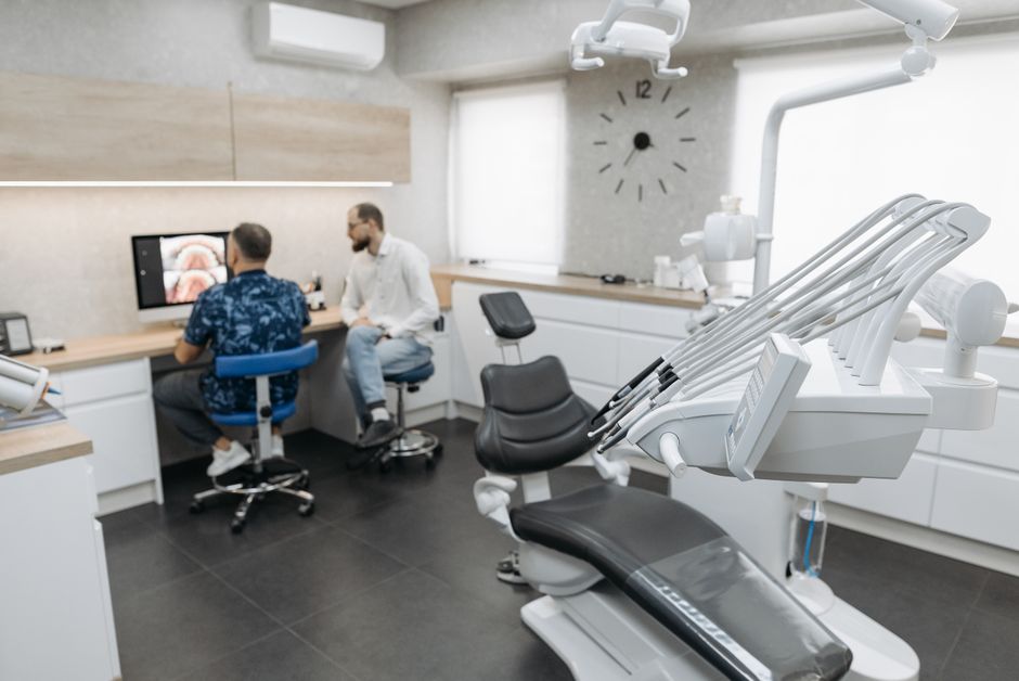
Digital dentistry: scanning, milling, and predictable lab communication
Intraoral scanners, digital articulators, and CAD/CAM workflows can improve data transfer and reduce distortion compared to conventional impressions—especially when margins are clearly visible and soft tissue management is strong. Digital tools also support patient communication through simulation and facilitate lab collaboration by standardizing forms, shade mapping, and reference images.
However, digital does not eliminate biological variables. Tissue inflammation, saliva control, and preparation clarity still determine the quality of the scan and the final fit. The most consistent outcomes come from combining digital tools with disciplined analog fundamentals.
Where veneers meet implant dentistry: full-arch and mixed cases
Many clinicians encounter patients who want a “smile makeover” but present with a mix of failing teeth, missing teeth, and periodontal compromise. In these cases, veneers may be part of a broader plan that includes implants or full-arch solutions. Understanding the planning hierarchy—prosthetic goal first, then surgical/restorative execution—helps avoid mismatched incisal planes, midlines, or occlusal schemes.
For clinicians exploring full-arch implant options, it can be helpful to review the prosthetic and stability rationale discussed in advantages of All-on-6 implants for full-arch predictability, especially when designing a stable occlusal platform that complements the patient’s aesthetic objectives.
Similarly, decision-making between different full-arch concepts requires careful consideration of bone availability, AP spread, prosthetic design, and maintenance. Our educational comparison of All-on-4 vs All-on-6 differences for full-arch implant planning can support structured thinking when veneers alone cannot meet the functional demands of a compromised dentition.
Common clinical pitfalls—and how training can reduce them
1) Over-preparation or under-preparation
Over-preparation compromises enamel bonding and may increase sensitivity risk; under-preparation often results in bulky contours and inflamed gingiva. Guided reduction using depth cuts and silicone indices helps keep preparations intentional and consistent.
2) Ignoring occlusion and parafunction
Veneer fractures and chipping are frequently linked to functional overload rather than “weak ceramics.” Aesthetic design should be coordinated with guidance, envelope of function, and protective strategies in at-risk patients.
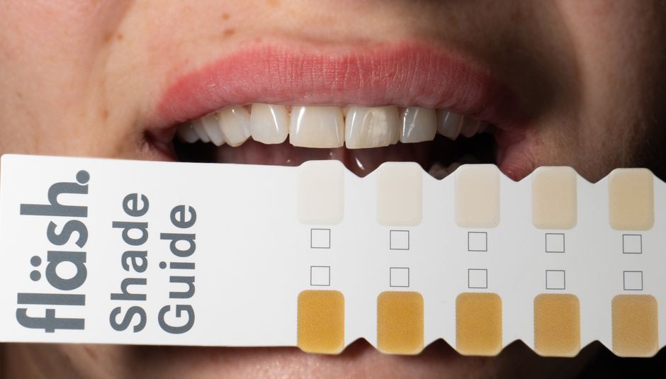
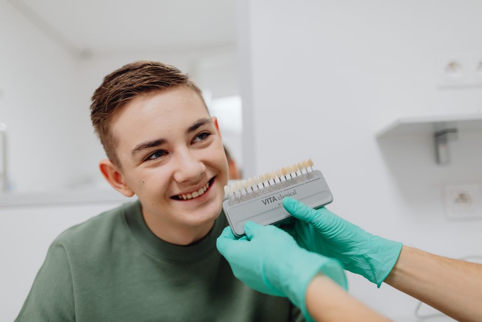
3) Inconsistent shade control
Shade is a system: lighting, photography, stump shade, ceramic thickness, and cement shade all contribute. Establishing a repeatable shade protocol with the lab is one of the most practical upgrades a clinician can make.
4) Cement remnants and soft tissue inflammation
Residual resin cement is a frequent cause of post-cementation inflammation. Good retraction, controlled tack-cure timing, and systematic cleanup protocols help protect the gingiva and preserve the final aesthetic result.
Learning veneer workflows at Istanbul Dental Academy
Because laminate veneers are technique-sensitive, many clinicians prefer to refine their skills through structured continuing dental education that combines theory with repeated hands-on practice. At Istanbul Dental Academy, our courses emphasize step-by-step clinical workflows—diagnostic records, mock-up driven preparation, digital planning integration, adhesive cementation protocols, and complication management—so participants can translate concepts into predictable chairside execution.
Hands-on training also supports interdisciplinary thinking: understanding when veneers are appropriate, when partial coverage restorations are a better option, and when implant-supported solutions should be considered as part of a comprehensive plan. This broader perspective is often what turns “good-looking” outcomes into stable, maintainable dentistry.
Conclusion
Porcelain laminate veneers can be a highly conservative and aesthetic solution when case selection is disciplined, enamel is preserved, and adhesive protocols are executed with precision. In minimal-invasive aesthetic dentistry, success is rarely about a single material choice—it is about an integrated workflow that respects biology, function, and communication.
This content is for educational purposes. Clinicians should evaluate each patient individually and follow current evidence, manufacturer instructions, and local regulations when planning and delivering care.
Diğer Yazılar