
BLOG
Microscope-Assisted Root Canal Shaping Techniques: A Clinical Education Guide
Blog Tarihi: 15/09/2026
Why Microscope-Assisted Shaping Matters in Modern Endodontics
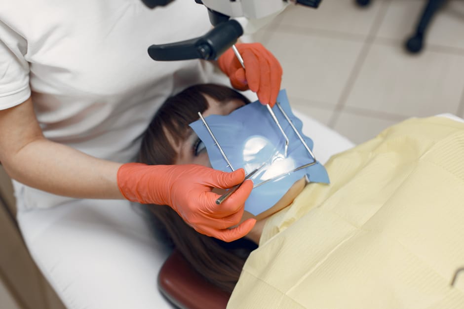
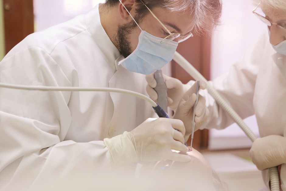
Root canal shaping is not simply “making space for irrigation and obturation.” It is a biologically driven, anatomy-respecting procedure in which the clinician must negotiate complex canal morphologies while preserving tooth structure and avoiding iatrogenic damage. The dental operating microscope (DOM) has moved from a “nice-to-have” tool to a core component of contemporary endodontic care in many clinics, primarily because it enhances visibility, supports finer motor control, and makes micro-anatomic details—such as calcifications, crack lines, accessory canal orifices, and subtle color changes in dentin—more clinically interpretable.
In Istanbul, where patients increasingly expect predictable, conservative treatments and high-quality restorations, microscope-assisted workflows also align with multidisciplinary dentistry. Endodontic precision affects the long-term success of restorative work, prosthodontic planning, and even implant decision-making—especially when clinicians are weighing tooth preservation against extraction and implant placement.
This content is for educational purposes and is not a substitute for diagnosis or treatment planning by a qualified clinician.
Optics, Ergonomics, and Workflow Setup
Magnification and illumination: the practical goal
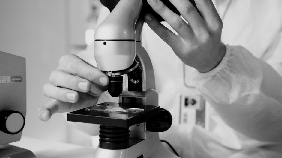
Microscope-assisted shaping is not about working at maximum magnification all the time. A practical approach is to use lower magnification for access refinement and coronal scouting, then increase magnification for locating orifices, verifying glide path patency, managing complex anatomy, and checking for procedural errors. Coaxial illumination is particularly helpful in deep access cavities and in teeth with restorative shadows, where headlight-based illumination may be insufficient.
Ergonomics: precision depends on posture
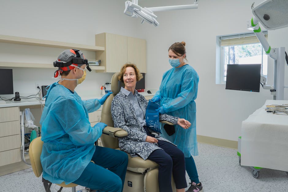
Many shaping errors originate from operator fatigue and poor posture. A stable seated position, neutral wrist alignment, and proper microscope positioning can improve tactile perception and reduce unintended file deflection. In hands-on training environments, clinicians often discover that their “usual” access angle changes under the microscope—sometimes for the better—because the DOM encourages a more centered approach to the pulpal floor.
Clinical Sequence: From Access to Glide Path Under the Microscope
Access cavity refinement: conservative yet functional
Microscope-assisted access is best viewed as anatomy-guided rather than simply “minimal.” The objective is to create straight-line access to the canal system while preserving pericervical dentin. Under magnification, clinicians can better differentiate pulpal floor anatomy, map dentin coloration, and recognize developmental grooves that suggest hidden anatomy. Ultrasonic tips, used carefully, can be more controlled under the DOM when troughing around calcified canal entries or removing dentin shelves.
Orifice location and canal scouting
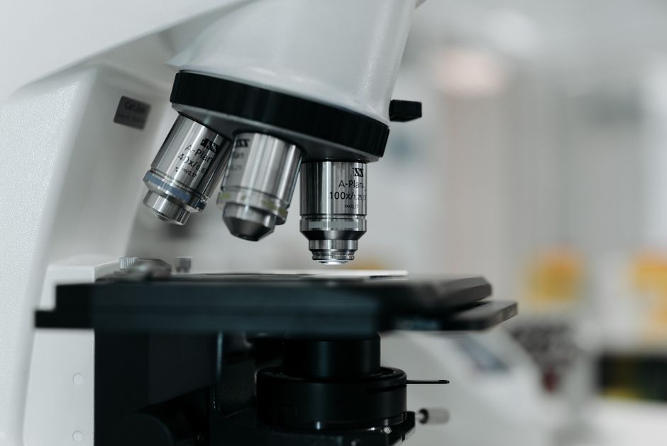
With improved illumination, orifice location becomes less of a “hunt” and more of a systematic exploration: visual cues (color, bleeding points, map lines), gentle ultrasonic troughing, and fine explorers or micro-openers. In calcified cases, the microscope helps the operator confirm that troughing is directed along the pulpal floor rather than into lateral dentin, reducing the risk of perforation.
Glide path: controlled, repeatable, and verified
A reproducible glide path reduces torsional stress on rotary/reciprocating files and supports centered shaping. Under the microscope, clinicians can confirm file entry, observe coronal file trajectory, and identify early warning signs of ledging or blockage. Whether using manual stainless-steel files, dedicated glide path rotary files, or a hybrid approach, the DOM supports the “look-and-feel” feedback loop that is essential for safe negotiation of narrow or curved canals.
Microscope-Assisted Shaping Techniques and Strategies
1) Crown-down vs. single-length: choosing a concept
Magnification does not replace a shaping philosophy—it enhances the clinician’s ability to execute it precisely. Crown-down strategies can reduce coronal interferences and improve irrigant penetration, while single-length strategies can offer procedural simplicity in selected anatomies. Under the DOM, the clinician can more confidently evaluate coronal enlargement, canal straight-line access, and whether the prepared shape remains conservative in critical dentin zones.
2) Curvature management and anti-transportation principles
Canal transportation is often invisible until it becomes clinically significant. Microscope-assisted inspection of the access trajectory, coronal third enlargement, and file path can reduce the likelihood of forcing instruments into curvature. A conservative coronal flaring approach—paired with a verified glide path—can help the clinician maintain a centered preparation and preserve apical anatomy.
3) Apical sizing: balancing debridement and preservation
Determining apical size is a biological and mechanical compromise. While larger apical sizes may facilitate irrigant exchange, overly aggressive shaping can weaken roots, increase the risk of procedural complications, and complicate obturation in curved canals. Under magnification, subtle signs—like a “shiny” canal wall indicating potential over-instrumentation or the presence of debris packing—can guide more informed decisions during instrumentation.
4) Irrigation and shaping: integrated, not sequential
Shaping is only one part of the cleaning objective. A microscope supports frequent canal inspection, patency checks, and debris management—particularly in cases where dentin mud obscures anatomy. While irrigation protocols depend on clinical judgment and case factors, the educational takeaway is that microscope use encourages clinicians to view shaping as a dynamic process: instrument, irrigate, recapitulate, reassess.
Common Pitfalls the Microscope Helps You Prevent (and Detect)
Ledges, blocks, and separated instruments
Early detection is the real advantage. Under the DOM, clinicians may notice subtle canal deviation, unusual file reflection, or debris accumulation that suggests impending blockage. While magnification does not eliminate instrument separation risk, it can facilitate prompt recognition and more controlled management pathways (e.g., improved visualization for ultrasonic staging in select situations).
Missed anatomy and untreated canals
Missed canals remain a frequent reason for endodontic failure. Magnification helps clinicians interpret the pulpal floor map, identify additional orifices, and inspect developmental grooves. This becomes especially relevant in maxillary molars, mandibular incisors, and premolars with variable canal systems, where “textbook anatomy” is the exception, not the rule.
Perforations and over-troughing
Perforation risk increases when access is performed without consistent reference to pulpal floor anatomy. The DOM improves depth perception and directional control, helping clinicians trough with greater precision. It also makes it easier to recognize anatomical danger zones early, before significant dentin removal occurs.
From Endodontics to Comprehensive Care: Why Shaping Precision Influences Other Specialties
Microscope-assisted shaping should be understood as a foundational skill that supports broader clinical goals. High-quality endodontics protects the restorative envelope, which is especially important when planning esthetic or functional rehabilitation.
Restorative and digital workflows: preserving tooth structure for predictability
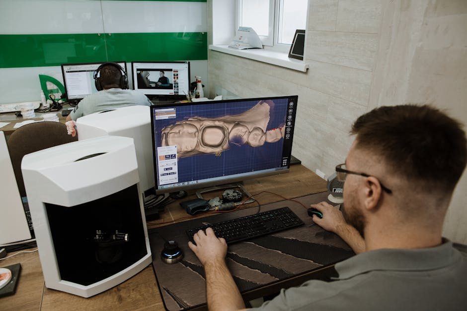
When a tooth is retained through well-executed endodontics, subsequent restorative decisions become more conservative and predictable. This same restorative mindset can be seen in digital planning and minimally invasive approaches discussed in Current Approaches to Managing Amelogenesis Imperfecta: A Modern Restorative and Digital Workflow, where material selection and digital tools are used to protect compromised tooth structure. While amelogenesis imperfecta is a distinct condition, the shared theme is clear: preserving and understanding hard tissue is central to long-term success.
Esthetic dentistry: endodontic foundations behind “smile makeovers”
Smile design often captures attention, but underlying endodontic and restorative integrity is what sustains outcomes. Cases involving discoloration, extensive restorations, or structural compromise may require careful endodontic assessment before veneers or full-coverage solutions are considered. For clinicians interested in the restorative-esthetic interface, Hollywood Smile: A Clinician’s Guide to Modern Smile Makeovers offers a broader look at modern planning—an area where endodontic precision quietly plays a critical supporting role.

Implant decision-making: saving teeth vs. replacing them
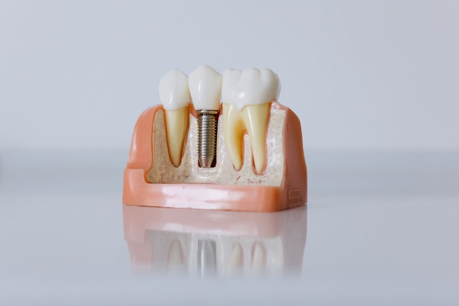
Microscope-assisted endodontics can influence case selection when clinicians and patients are weighing tooth retention against extraction and implant placement. Implant therapy can be highly successful, but it requires careful selection and planning. For a balanced educational overview of implant timelines and indications, consider reading Same-Day Dental Implants: Advantages, Limitations, and Case Selection and Is One-Day Dental Implant Treatment Really Possible?. Understanding these pathways helps clinicians communicate options transparently while keeping endodontic excellence as a viable tooth-preservation strategy.
Prosthodontic outcomes: precision upstream affects passive fit downstream
Although passive fit is typically discussed in implant prosthodontics, the broader principle applies across dentistry: accuracy earlier in the workflow reduces complications later. For implant-supported restorations, Achieving Passive Fit in Implant-Supported Prostheses: Clinical & Lab Strategies highlights how clinical and laboratory steps combine to improve predictability—mirroring how endodontic shaping, disinfection, and obturation must integrate as one coherent system.
Training Microscope-Assisted Shaping: What to Practice in Hands-On Education
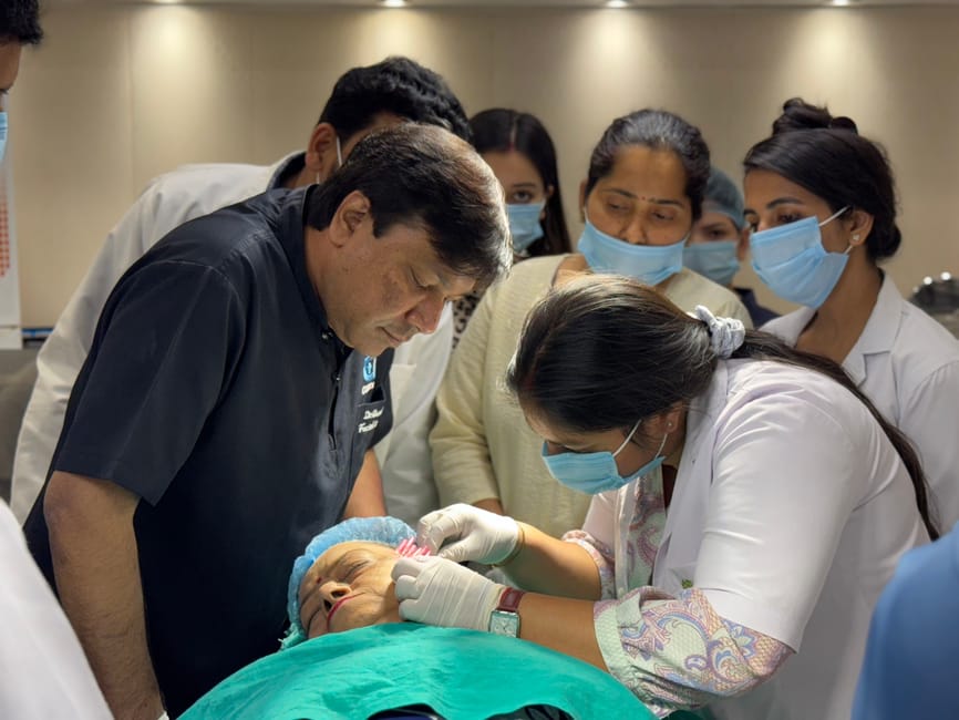
Many clinicians first encounter the DOM as an “advanced tool,” but proficiency is built through structured repetition. At Istanbul Dental Academy, microscope-based training is approached as a practical workflow skill: positioning, access refinement, canal negotiation, glide path confirmation, and shaping verification under magnification—followed by case-based discussions on decision-making and complication prevention.
In hands-on settings, participants typically benefit from:
• Ergonomic coaching (chair position, microscope alignment, assistant coordination)
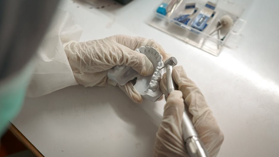
• Access design exercises on extracted teeth or training models with realistic anatomy
• Glide path and shaping drills emphasizing patency, recapitulation, and controlled instrumentation
• Visual checkpoints for detecting iatrogenic risk early (transportation, ledges, over-preparation)
The goal is not to memorize a single “best” system, but to develop a repeatable method that adapts to canal anatomy and integrates with restorative planning.
Key Takeaways
Microscope-assisted root canal shaping is a practical, skill-based upgrade that supports safer access, more controlled glide path creation, improved anatomy detection, and earlier recognition of procedural risks. For dental professionals pursuing continuing education in Istanbul, the DOM can be a meaningful bridge between endodontic fundamentals and modern multidisciplinary dentistry—where restorative, digital, esthetic, and implant workflows increasingly intersect.
This content is for educational purposes. Clinical decisions should be made based on individual patient factors, current evidence, and professional guidelines.
Diğer Yazılar