BLOG
Bleeding Gums: What Conditions Could They Signal? A Dental Education Guide
Blog Tarihi: 14/06/2026

Bleeding gums: a small sign with a wide differential
Gingival bleeding—whether it happens during brushing, flossing, or spontaneously—often reflects inflammation within the periodontal tissues. In daily practice, it is one of the most frequent patient complaints, yet it can be underestimated because it may be painless and intermittent. From an educational perspective, bleeding gums are best approached as a clinical sign rather than a diagnosis, prompting a structured evaluation of local plaque-related disease, iatrogenic factors, patient habits, and systemic conditions.
This content is for educational purposes and aims to support clinical thinking for dental professionals, dentists, and students. If a patient presents with persistent or unexplained bleeding, clinicians should consider comprehensive periodontal and medical assessment and coordinate care when appropriate.
Common local causes: gingivitis and periodontitis
Plaque-induced gingivitis
The most common cause of bleeding on probing (BOP) and home-care bleeding is plaque-induced gingivitis. Biofilm accumulation at the gingival margin triggers an inflammatory response, increasing vascular permeability and making tissues more prone to bleed with minor trauma. Clinically, gingivitis may present with erythema, edema, loss of stippling, and BOP, often without attachment loss.
Because gingivitis is typically reversible with effective plaque control, clinician communication matters: patients may normalize bleeding for years. In education and patient-centered care, it helps to frame bleeding as an “early warning light” and to connect home-care techniques with measurable parameters such as plaque score and BOP reduction.
Periodontitis (and why bleeding should not be ignored)
When inflammation persists and the host response, microbial dysbiosis, and risk factors align, gingivitis may progress to periodontitis—characterized by clinical attachment loss, pocket formation, and radiographic bone loss. Bleeding can be present, but notably, some advanced cases may bleed less due to fibrotic tissue changes or smoking-related vasoconstriction. That is why a thorough periodontal charting and radiographic evaluation remain essential.
For dentists involved in implant and restorative workflows, periodontal stability is not an optional “extra.” In contemporary treatment planning—especially for esthetic restorations and implants—periodontal diagnosis directly influences prognosis, maintenance intervals, and long-term outcomes.
Mechanical and iatrogenic contributors
Aggressive brushing, traumatic flossing, and device misuse
Patients often attribute bleeding to “brushing too hard,” and while mechanical trauma can contribute, persistent bleeding usually indicates underlying inflammation. Clinically, it is helpful to evaluate brush type, technique, interdental aids, and patient dexterity. Overzealous flossing can cause papillary trauma, while underuse can allow interdental biofilm to mature—both scenarios may present as bleeding.
Overhanging margins, open contacts, and restorative design
Iatrogenic plaque-retentive factors—overhanging restorations, poor emergence profile, subgingival margin placement without adequate biologic width considerations, and open contacts—can sustain inflammation and bleeding. From a restorative and prosthodontic viewpoint, gingival health should be assessed before definitive impressions, scans, and cementation steps.
Digital workflows can support better marginal integrity and contour control when used appropriately. For example, students exploring implant-supported restorations can benefit from understanding how a digital design translates into soft-tissue response and cleansability, as discussed in CAD/CAM technology in modern implant dentistry and the digital workflow.
Gum bleeding and gingival recession: an important overlap
Gingival recession does not automatically cause bleeding, but recession-prone sites may be associated with inflammation, thin phenotype, traumatic brushing, or malpositioned teeth. Recession also complicates plaque control by increasing sensitivity and making patients avoid brushing the area—potentially increasing inflammation and bleeding.
When clinicians see bleeding concentrated around recession defects, it can be a cue to assess phenotype, frenal pull, traumatic occlusion, and the patient’s hygiene technique. Treatment planning may involve non-surgical approaches (behavior change, professional debridement, inflammation control) and, when indicated, mucogingival surgery. For an evidence-informed overview, see gum recession treatment options—surgical and non-surgical approaches in modern dentistry.
Systemic and lifestyle factors that may amplify gingival bleeding
Smoking and nicotine products
Smoking is strongly associated with periodontitis, yet paradoxically, smokers may exhibit reduced visible bleeding due to vasoconstrictive effects. This can mask disease severity. For clinicians, this means bleeding is not the only indicator—probing depth, attachment levels, radiographs, and risk profiling remain essential.
Diabetes and glycemic control
Diabetes is a major risk factor for periodontal disease. Hyperglycemia can alter host response, collagen metabolism, and wound healing, potentially increasing susceptibility to inflammation and bleeding. Clinically, periodontal findings may be an opportunity to encourage interprofessional collaboration and to emphasize maintenance and risk reduction. The relationship also matters in implant dentistry: in patients with diabetes, evaluating periodontal status and glycemic control is part of responsible planning. For further clinical discussion, explore dental implant success in patients with diabetes and what clinicians should know.
Hormonal changes: pregnancy, puberty, and oral contraceptives
Hormonal fluctuations can modulate gingival inflammatory response to plaque, leading to pregnancy gingivitis or puberty-associated gingival changes. Bleeding may increase even with similar plaque levels. The key educational message is that plaque remains the primary trigger, while hormones can heighten the response—reinforcing the importance of tailored preventive strategies and frequent professional monitoring.
Medications and bleeding tendency
Certain medications can influence gingival tissues or bleeding risk. Anticoagulants and antiplatelet agents may increase bleeding during brushing or after instrumentation; calcium channel blockers and other drugs may contribute to gingival overgrowth, which can trap plaque and exacerbate inflammation. Clinicians should obtain a thorough medical history and coordinate with the patient’s physician when necessary, particularly before surgical periodontal or implant procedures.
Nutritional factors and hematologic conditions
Although less common than plaque-induced disease, nutritional deficiencies (e.g., vitamin C deficiency) and hematologic disorders can present with gingival bleeding. Red flags may include spontaneous bleeding, petechiae, bruising, fatigue, or bleeding disproportionate to local inflammation. In such cases, dental findings can be an important prompt for medical referral and appropriate laboratory evaluation.
When bleeding gums may signal deeper dental pathology
Endodontic-periodontal interactions
Localized bleeding and swelling around a specific tooth may be linked to periodontal pockets, but clinicians should also consider endodontic pathology and endo-perio lesions. Accurate diagnosis relies on percussion, palpation, pulp vitality testing, probing patterns, and radiographic interpretation. Visualization can become especially important in retreatment scenarios, cracked teeth assessments, or locating missed anatomy.
For clinicians refining diagnostic precision, the use of magnification is increasingly central to modern practice. Istanbul Dental Academy highlights clinical visibility and ergonomics in advanced training; related reading includes the role of the dental operating microscope in modern endodontics for better vision and outcomes.
Peri-implant mucositis and peri-implantitis
Bleeding around implants—especially bleeding on gentle probing—may indicate peri-implant mucositis, which can progress to peri-implantitis if inflammation and bone loss develop. Because implants lack a periodontal ligament and have different soft-tissue attachment characteristics, clinical monitoring should be systematic, including baseline probing depths, radiographs, and maintenance protocols.
Patients may interpret peri-implant bleeding as “normal” irritation, so education is essential. Clinicians should evaluate plaque control, restoration contours, and occlusal factors, and ensure the patient is enrolled in supportive peri-implant care.
Clinical assessment: a structured approach for dentists and students
History and risk profiling
A focused history helps differentiate transient trauma from ongoing inflammation. Key points include onset, frequency, oral hygiene routine, last prophylaxis, smoking status, systemic diseases (especially diabetes), medications, pregnancy status, and previous periodontal therapy. Risk profiling is not merely administrative; it informs prognosis, recall intervals, and patient communication.
Examination: indices and periodontal charting
At minimum, clinicians should record plaque indices, BOP, probing depths, recession, clinical attachment level when indicated, furcation involvement, mobility, and mucogingival considerations. Radiographs should be interpreted in light of clinical data; bone levels without probing data can mislead, and vice versa.
Documentation and communication
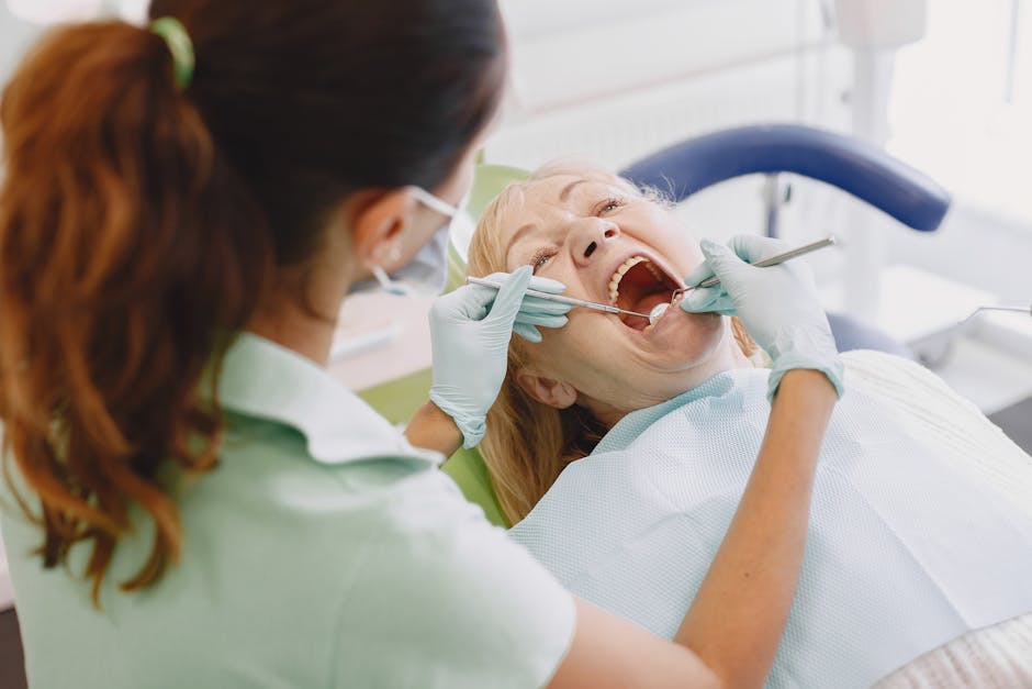
Bleeding is a valuable educational tool: showing patients bleeding sites during probing can motivate behavior change when explained respectfully. Dental photography and intraoral scanning can also support monitoring and case presentation, helping patients visualize inflammation and understand the link between daily habits and clinical outcomes.
Why periodontal health matters for esthetics, restorations, and implants
Bleeding gums are not only a periodontal concern—they can compromise restorative margins, impression accuracy (analog or digital), adhesive protocols, and esthetic outcomes in smile design. Inflamed tissue can alter gingival contours and papillae, increasing the risk of black triangles or asymmetry after veneers and crowns. For clinicians, periodontal stabilization is often the prerequisite for predictable cosmetic dentistry.
In implant dentistry, soft-tissue management and maintenance are equally critical. Patients may ask about accelerated solutions, including immediate loading concepts. While same-day protocols can be appropriate in selected cases with careful planning, they require rigorous diagnosis, risk assessment, and workflow coordination. For an educational overview, read whether one-day dental implant treatment is really possible.
Training perspective: building competence from diagnosis to hands-on care
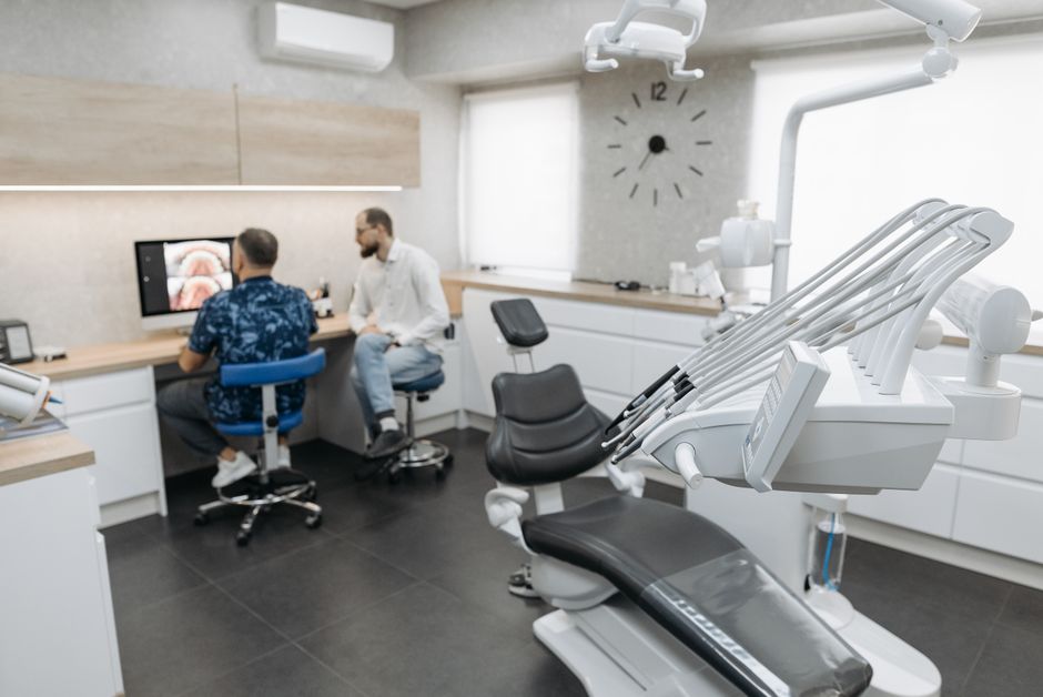
For dental professionals, managing gingival bleeding effectively is less about a single technique and more about integrating diagnosis, periodontal fundamentals, restorative design principles, and patient communication. At Istanbul Dental Academy, our continuing dental education approach emphasizes bridging theory with clinical implementation—supporting dentists and students who want to sharpen periodontal assessment, improve treatment planning for restorative and implant cases, and practice hands-on workflows in a structured learning environment.
Whether your focus is periodontology, restorative dentistry, endodontics, or implant prosthodontics, a consistent takeaway remains: bleeding gums are a sign worth investigating systematically, documenting carefully, and using as an opportunity to improve overall oral health outcomes through evidence-based, patient-specific care.
Key takeaways
Bleeding gums most often indicate plaque-induced inflammation, but they can also be influenced by iatrogenic factors, systemic conditions (notably diabetes), hormonal changes, medications, and less common hematologic or nutritional issues. A structured assessment—history, periodontal charting, and appropriate imaging—helps clinicians identify risk, establish diagnosis, and plan care that supports long-term periodontal and peri-implant stability.
Diğer Yazılar