BLOG
The Role of CAD/CAM Technology in Modern Implant Dentistry
Blog Tarihi: 14/06/2026

Why CAD/CAM Matters in Implant Dentistry
CAD/CAM (computer-aided design and computer-aided manufacturing) has moved from being a “nice-to-have” technology to a central pillar of modern implant workflows. For dental professionals, its value is not simply speed or aesthetics; it is the ability to standardize steps, reduce analog errors, and create a more traceable, data-driven clinical pathway from diagnosis to final prosthesis.
In Istanbul—where patient expectations are high and complex rehabilitations are common—digital workflows can also support communication across teams (surgeon, restorative dentist, lab technician) and across disciplines such as periodontology, prosthodontics, and restorative dentistry. At Istanbul Dental Academy, digital dentistry is approached as a clinical skillset rather than a software demonstration: clinicians learn to integrate scanning, design, and manufacturing decisions with biological principles and risk management. This content is for educational purposes and does not replace individualized clinical judgment.
Core Components of a CAD/CAM Implant Workflow
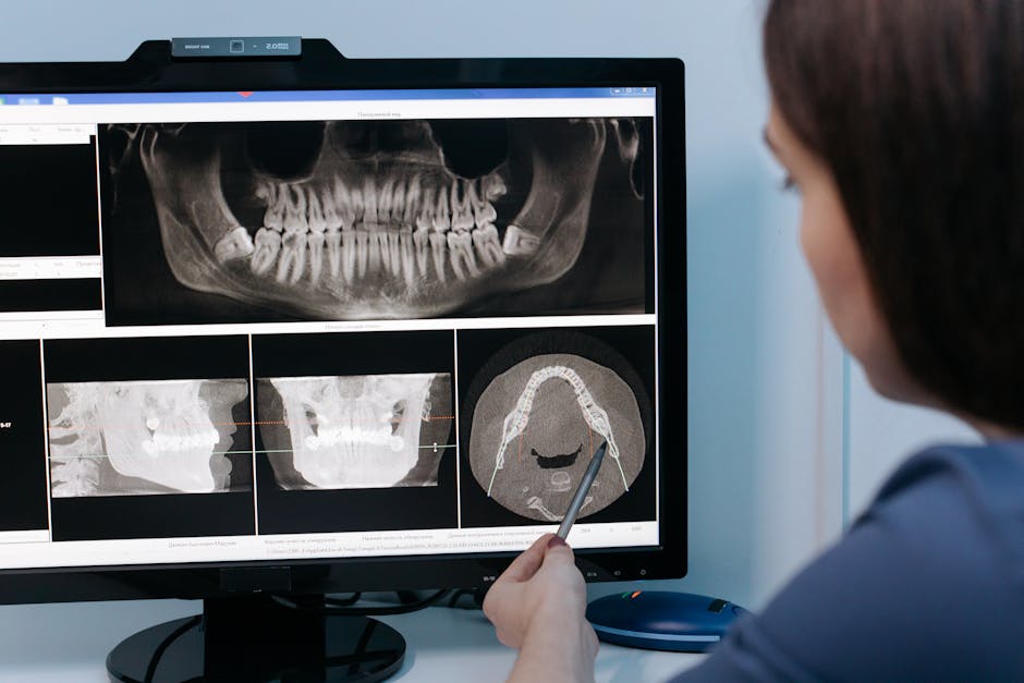
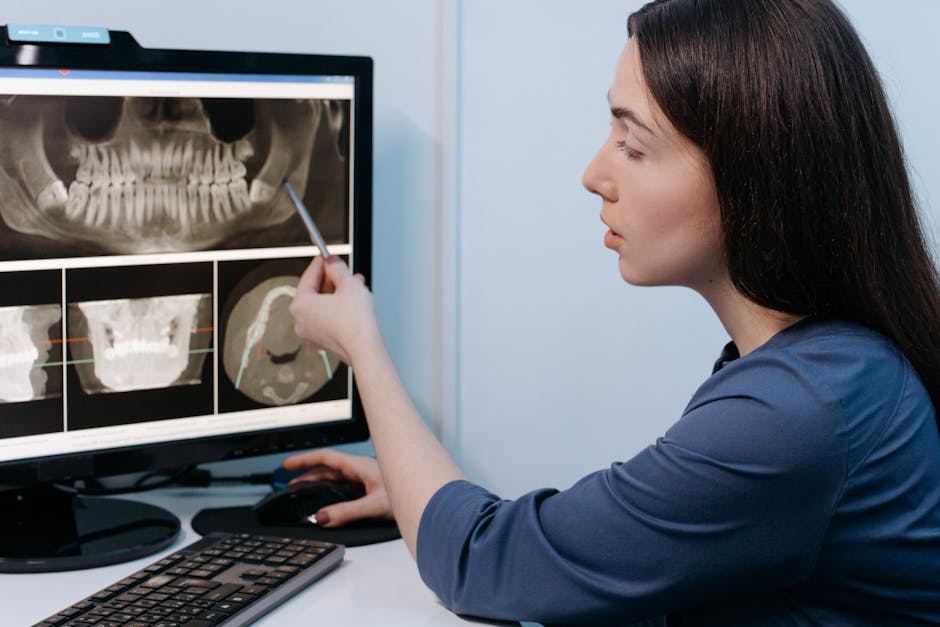
1) Data capture: intraoral scanning and CBCT integration
Implant treatment is information-sensitive. CAD/CAM begins with accurate data acquisition: intraoral scans capture soft-tissue contours, occlusal relationships, and existing restorations; CBCT provides three-dimensional bone anatomy and proximity to critical structures. When these datasets are merged, the clinician can plan prosthetically driven implant positions rather than placing implants based solely on available bone.
This digital diagnostic mindset connects naturally to broader “digital smile” expectations. When patients ask for a comprehensive aesthetic upgrade, implant planning often overlaps with smile design decisions, such as midline, occlusal plane, and tooth proportions—topics explored in descriptive guidance on Hollywood Smile techniques, materials, and clinical workflow. Even when implants are not placed in the aesthetic zone, understanding the aesthetic blueprint helps ensure restorations harmonize with the overall facial and dental composition.
2) Virtual implant planning: prosthetic-driven positioning
CAD planning platforms allow clinicians to select implant type and diameter virtually, simulate angulation, evaluate restorative space, and plan for screw-retained or cement-retained designs. Prosthetic-driven planning generally aims to align implant position with the final crown’s emergence profile and occlusal scheme, supporting hygiene access and reducing risk of prosthetic complications.
In educational settings, planning is often where clinicians discover how small changes in angulation can alter restorative outcomes. A key learning point is that “ideal” positioning is a balance between anatomy, prosthetic requirements, and tissue stability—especially in the anterior region where papilla support and emergence profile are critical.
3) Guided surgery and verification
Once planning is complete, a surgical guide can be designed and manufactured. Guides may be tooth-supported, mucosa-supported, or bone-supported, and their selection depends on stability, access, and clinical scenario. When used appropriately, guided surgery can help translate the virtual plan into the mouth with improved consistency—particularly for multiple implants, limited interarch space, or proximity to anatomical structures.
Guided workflows are also relevant in time-sensitive protocols. For clinicians exploring accelerated pathways, it is useful to contextualize digital planning with extraction and immediate placement protocols, as discussed in Same-Day Tooth Extraction and Immediate Implant Placement: a clinical guide. While digital tools can improve planning accuracy, case selection and tissue management remain decisive.
4) CAD/CAM prosthetics: from abutments to full-arch solutions
On the restorative side, CAD/CAM supports fabrication of custom abutments, crowns, bridges, and full-arch frameworks. Compared with purely conventional steps, digitally designed components can improve fit, streamline remakes, and provide repeatable parameters (cement space, connector dimensions, emergence profile).
Material selection is central here. Zirconia, titanium, cobalt-chromium, hybrid ceramics, and lithium disilicate may all play roles depending on location, load, and aesthetic needs. In implant prosthodontics, clinicians must also consider screw channel positioning, retrievability, and occlusal design to mitigate complications such as screw loosening or ceramic chipping.
Clinical Advantages—and the “Hidden” Limitations
Precision and traceability
One of CAD/CAM’s strongest advantages is traceability: the clinician can document initial scans, the planned implant position, guide design parameters, and final restoration design. This not only supports team communication but can also make follow-up assessments more objective when managing complications or monitoring tissue changes over time.
Reduced analog distortion (but not reduced responsibility)
Digital impressions can reduce inaccuracies associated with impression materials, tray selection, and stone model expansion. However, scanning errors can still occur—particularly around subgingival margins, reflective surfaces, or mobile soft tissues. “Digital” does not automatically mean “accurate”; it means the accuracy depends on technique, calibration, and verification.
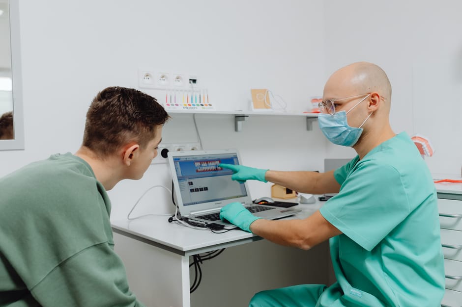
Efficiency and patient experience
Shorter chair time, fewer appointments, and a more visual communication style can improve the patient experience. In complex cases, showing a patient a proposed digital wax-up or a 3D plan can enhance understanding and consent quality. Still, clinicians should avoid overpromising speed; biological healing timelines and tissue stability remain fundamental constraints.
Where CAD/CAM does not replace fundamentals
CAD/CAM cannot compensate for weak diagnosis, compromised periodontal health, or inadequate occlusal planning. For example, uncontrolled inflammation or poor plaque control can destabilize soft tissue outcomes and increase the risk of peri-implant complications. A strong periodontal baseline is essential; clinicians may find it useful to revisit foundational principles in clinical insights on gum disease, early signs, and risk factors when assessing implant candidates or planning long-term maintenance.
Soft-Tissue Management in the Digital Era
Implant success is not only osseointegration; it is also the stability of the peri-implant mucosa, the emergence profile, and the patient’s ability to maintain hygiene. CAD/CAM supports this through emergence profile design and provisionalization strategies, but soft-tissue biology still requires hands-on clinical competence.
Designing emergence profiles and provisional restorations
Digitally designed provisionals can help shape peri-implant soft tissue, especially in the aesthetic zone. Proper contouring should support the tissue without excessive pressure, and designs should be evaluated in the mouth for phonetics, cleansability, and occlusion. Digital libraries help standardize abutment geometries, but individualized contouring remains critical.
Grafting and biomaterials considerations
When volume deficiencies exist, clinicians may consider soft-tissue augmentation or ridge preservation depending on the case. Modern biomaterials are frequently discussed in continuing education because they influence aesthetic stability and patient comfort. For those exploring grafting adjuncts, Asellüler Dermal Matriks: its use in dentistry provides educational context on one category of biomaterial used in soft-tissue procedures. The decision to use any grafting approach should be based on case assessment, operator experience, and current evidence.
Interdisciplinary Links: Endodontics, Restorative Dentistry, and Digital Precision
Implant dentistry often intersects with endodontic decision-making and restorative planning—especially in borderline cases where tooth preservation may still be possible. Digital dentistry does not end at implants; it also supports minimally invasive restorative workflows, post-endodontic restorations, and precision evaluation of margins and anatomy.
Magnification and illumination are important complements to digital planning. While CAD/CAM enhances design and manufacturing, the clinical steps—caries removal, access cavity refinement, adhesive isolation, margin finishing—still depend on visibility and operator control. Clinicians seeking a broader view of precision-enhancing tools may also benefit from Dental Operating Microscope in modern endodontics: better vision and better outcomes, which highlights how enhanced visualization can support predictable clinical execution across disciplines.
From Single Crowns to Full-Arch: How CAD/CAM Scales
Single-tooth implants
For single units, CAD/CAM helps with accurate occlusal morphology, proximal contacts, and emergence profile customization. Digital workflows can also streamline the creation of implant crowns that integrate well with adjacent teeth—particularly important in the aesthetic zone.
Multiple implants and bridges
As case complexity increases, digital planning and manufacturing can reduce cumulative inaccuracies. Framework design parameters (connector size, pontic design, implant angulation compensation) can be standardized and communicated clearly to the laboratory team.
Full-arch rehabilitation
Full-arch implant therapy is where the advantages of CAD/CAM become highly visible: controlled prosthetic setup, verification strategies, and the ability to reproduce prostheses if repairs are needed. Nonetheless, full-arch treatment also magnifies risks—errors in vertical dimension, occlusal scheme, or implant distribution can have large consequences. For this reason, hands-on training and supervised case discussions are particularly valuable before implementing full-arch protocols in daily practice.
How to Build Competence: Training Beyond the Software
Digital implant dentistry is often marketed as a technological upgrade, but in clinical reality it is a workflow upgrade. Clinicians must learn how to:
• Evaluate scan quality and recognize artifacts
• Merge datasets accurately and verify reference points
• Plan implants prosthetically while respecting anatomy
• Select guide type and understand tolerance/offset concepts
• Design provisional and definitive restorations with cleansability in mind
• Communicate effectively with the lab using digital prescriptions
Istanbul Dental Academy emphasizes continuing dental education that integrates these digital steps with clinical fundamentals—occlusion, periodontal assessment, soft-tissue management, and complication prevention. Hands-on components are essential because many “digital errors” are actually clinical errors (unstable scan bodies, inadequate retraction, poor isolation, improper verification).
Practical Takeaways for Daily Practice
CAD/CAM technology can support more predictable implant outcomes by improving planning, enhancing team communication, and enabling precise prosthetic fabrication. However, the best results typically come from clinicians who pair digital competence with strong biological understanding—periodontal health, soft-tissue stability, and occlusal control remain central.
For dental professionals in Istanbul and international clinicians training in the city, adopting CAD/CAM is often less about purchasing devices and more about building a reliable clinical sequence—one that can be repeated, audited, and refined over time. This content is for educational purposes and is not a substitute for diagnosis or treatment planning for any individual patient.
Diğer Yazılar


