BLOG
Bone Augmentation Before Implant Placement in Atrophic Jaws: A Clinical Education Guide
Blog Tarihi: 25/06/2026
Why atrophic jaws matter in implant dentistry
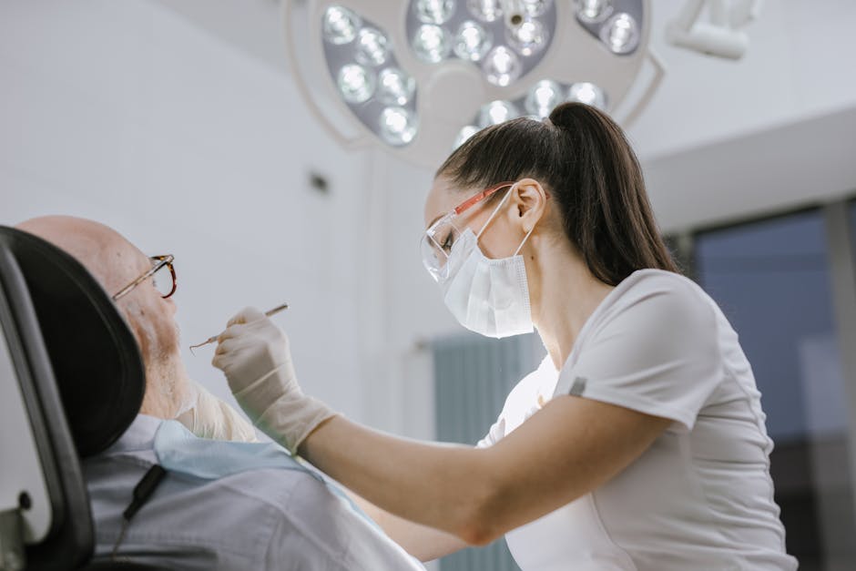
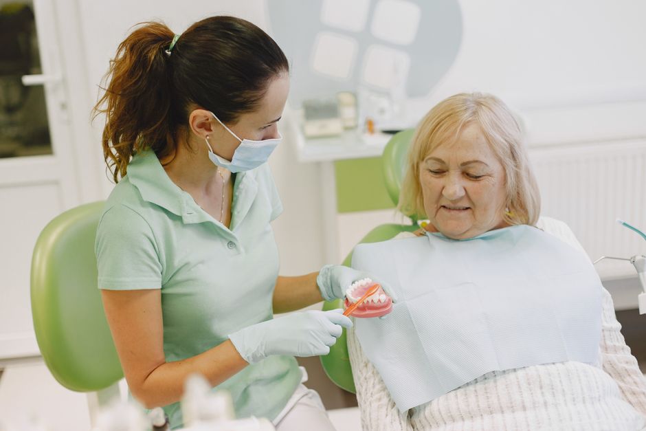
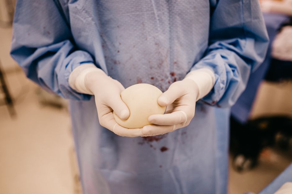
Tooth loss can trigger progressive alveolar bone resorption, and long-term edentulism may leave clinicians with limited ridge width, reduced vertical height, and compromised soft-tissue architecture. These changes define what many clinicians describe as an “atrophic jaw”—a clinical reality that can complicate implant positioning, primary stability, prosthetic emergence profiles, and long-term peri-implant health. In educational settings, atrophic cases are often where planning discipline becomes most visible: the clinician must balance restorative goals with anatomical limitations, risk management, and surgical execution.
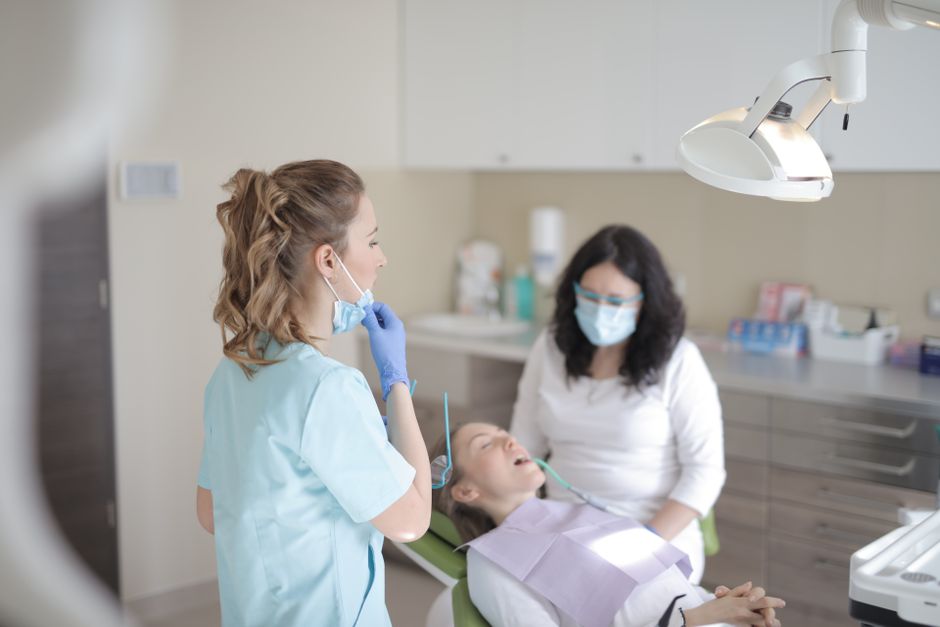
At Istanbul Dental Academy, we approach atrophic jaw rehabilitation as a team-based workflow—prosthodontically driven planning, periodontology-informed soft-tissue management, and oral surgery technique—supported by hands-on training. This article is for educational purposes and aims to provide a structured overview of bone augmentation options commonly considered before or alongside implant placement.
Diagnosis first: defining the defect and the prosthetic goal
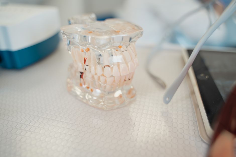
Before choosing an augmentation strategy, clinicians should clarify two questions: (1) What is missing—width, height, or both? (2) Where should the implant ideally be positioned to support the definitive restoration? In contemporary implant dentistry, a prosthetically driven plan often reduces “compromise implants” that later force bulky contours, hygiene challenges, or unfavorable load directions.
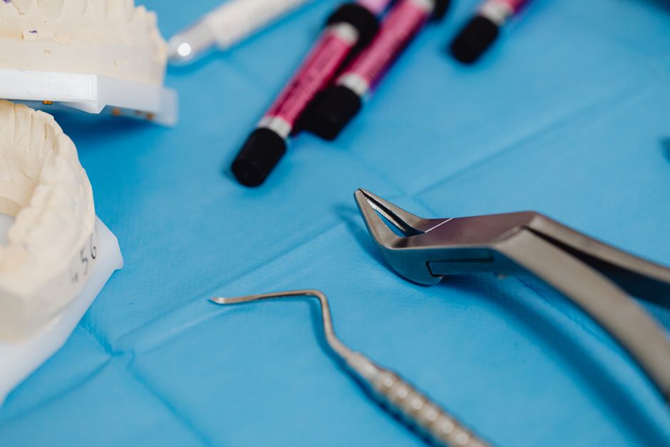
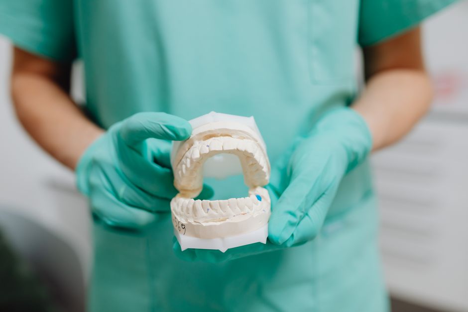
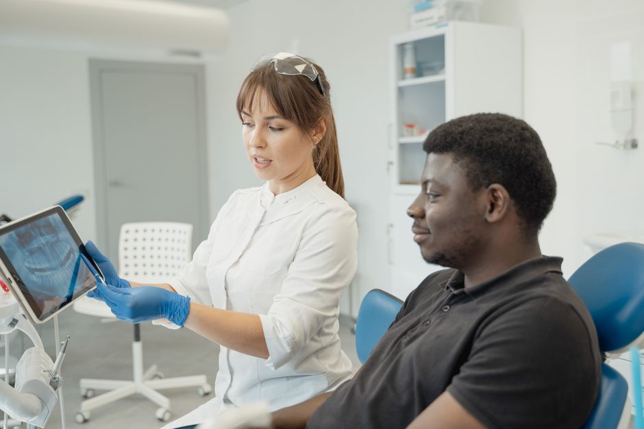
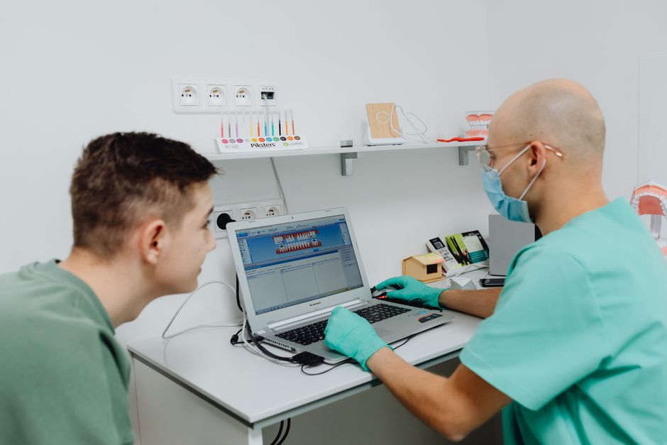
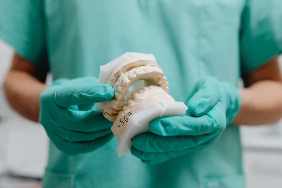
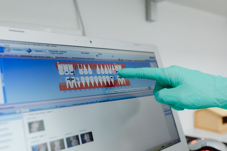
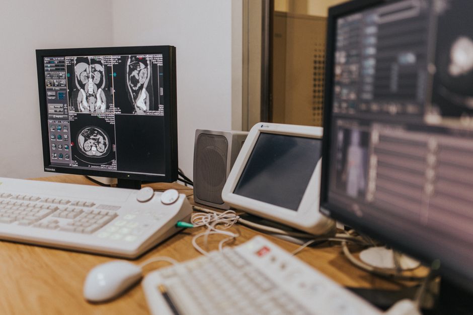
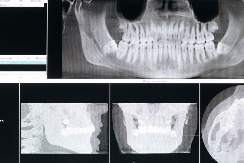
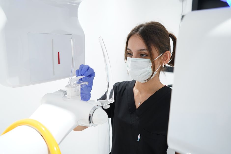
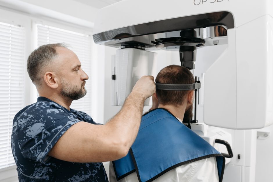
Clinical examination and intraoral photography support baseline documentation, while radiographic analysis—often via CBCT—helps evaluate ridge morphology, sinus/nasal floor proximity, mandibular canal position, and the quality/quantity of available bone. Digital dentistry tools can integrate DICOM and intraoral scan data to simulate implant placement and guide augmentation volumes when indicated.
It can be helpful to contextualize augmentation needs within aesthetic priorities. For example, anterior cases may demand ridge volume that supports papillae and natural emergence, similar in philosophy to aesthetic planning methods used in smile design. If you’re building a comprehensive treatment-planning mindset, consider how facial and dental proportions guide restorative outcomes in face-shape–oriented smile design with digital planning—the same “start with the end” logic applies to implant restorations in atrophic ridges.
Common clinical patterns of atrophy
Horizontal deficiency (narrow ridge) is frequently encountered, especially after extraction without ridge preservation. Vertical deficiency (reduced height) may result from long-standing resorption, periodontal history, or trauma and often carries higher surgical complexity and complication risk. Combined defects demand careful staging and may require advanced grafting, customized membranes/meshes, or alternative implant strategies.
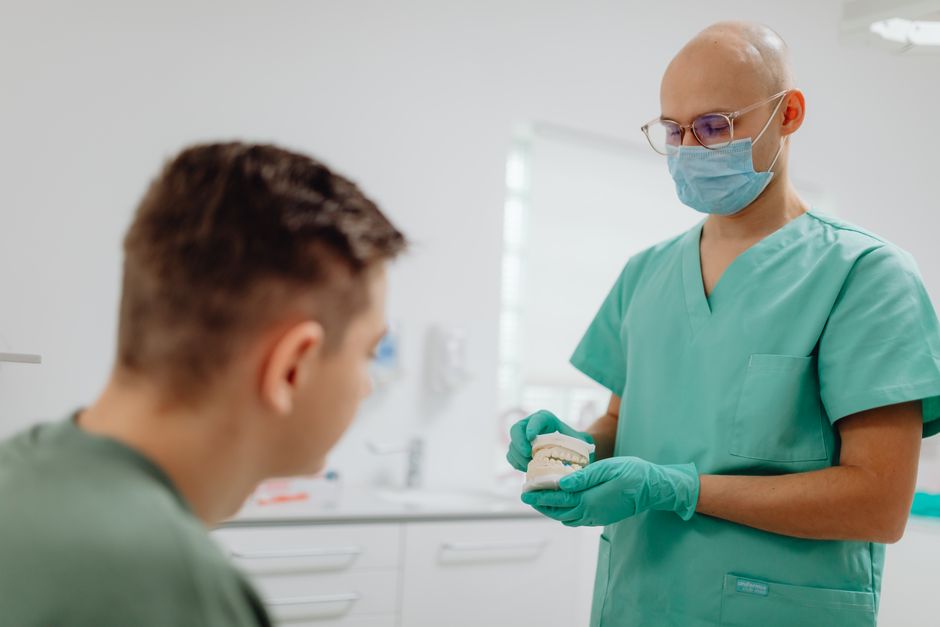
Bone augmentation options: an educational overview
Augmentation is not a single technique but a spectrum. Case selection is influenced by defect morphology, soft-tissue quality, systemic and local risk factors, patient expectations, and clinician experience. Below are common approaches discussed in structured implant education.
Guided bone regeneration (GBR)
GBR is widely used for localized horizontal defects and certain contained vertical components. The principle is straightforward: a barrier membrane supports space maintenance while particulate graft material and clot stabilization encourage bone fill. In practice, outcomes depend on flap design, tension-free closure, membrane selection (resorbable vs non-resorbable), and strict control of micromovement and contamination.
From a training perspective, GBR is an excellent framework for learning fundamentals—incision planning, periosteal release, stabilization (tacks/screws), and suturing—because small lapses in soft-tissue management may lead to membrane exposure and graft loss. Hands-on courses can accelerate competence by allowing clinicians to repeat these steps with faculty feedback.
Ridge split / ridge expansion
When the ridge is narrow but has adequate height and favorable bone quality, ridge splitting or expansion can sometimes create implant space while minimizing graft volume. Educationally, the technique emphasizes careful manipulation of cortical plates, controlled osteotomies, and an understanding of bone elasticity—often more predictable in maxillary bone than mandibular bone.
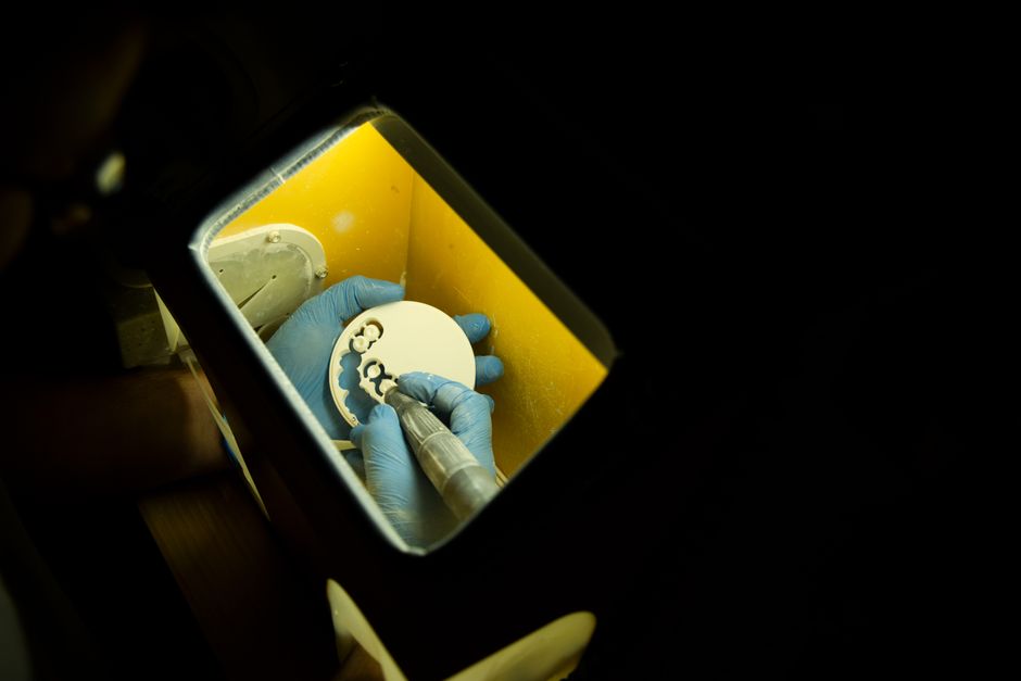
Block grafting (autogenous or allogenic blocks)
Block grafting may be considered for more significant horizontal deficiencies or when space maintenance is challenging. Autogenous blocks can provide osteogenic potential but introduce donor-site morbidity; allogenic blocks may reduce morbidity but require meticulous adaptation, fixation, and soft-tissue coverage. In teaching settings, block grafting is often positioned as an advanced module because success relies on stable fixation, intimate contact, and careful tissue handling.
Sinus floor elevation (lateral or transcrestal)
Posterior maxillary atrophy commonly intersects with pneumatized sinuses. Sinus augmentation can create vertical bone height for implant placement, either staged or simultaneous depending on residual bone height and primary stability goals. Education here focuses on anatomy, membrane management, and complication handling (e.g., Schneiderian membrane perforation).
Vertical ridge augmentation and advanced reconstruction
True vertical augmentation—using GBR with non-resorbable membranes, titanium meshes, or staged approaches—can be more technique-sensitive and complication-prone than horizontal augmentation. Alternative strategies may include short implants, angled implants, or prosthetic redesign, but these choices should be framed within biomechanical and hygiene considerations.
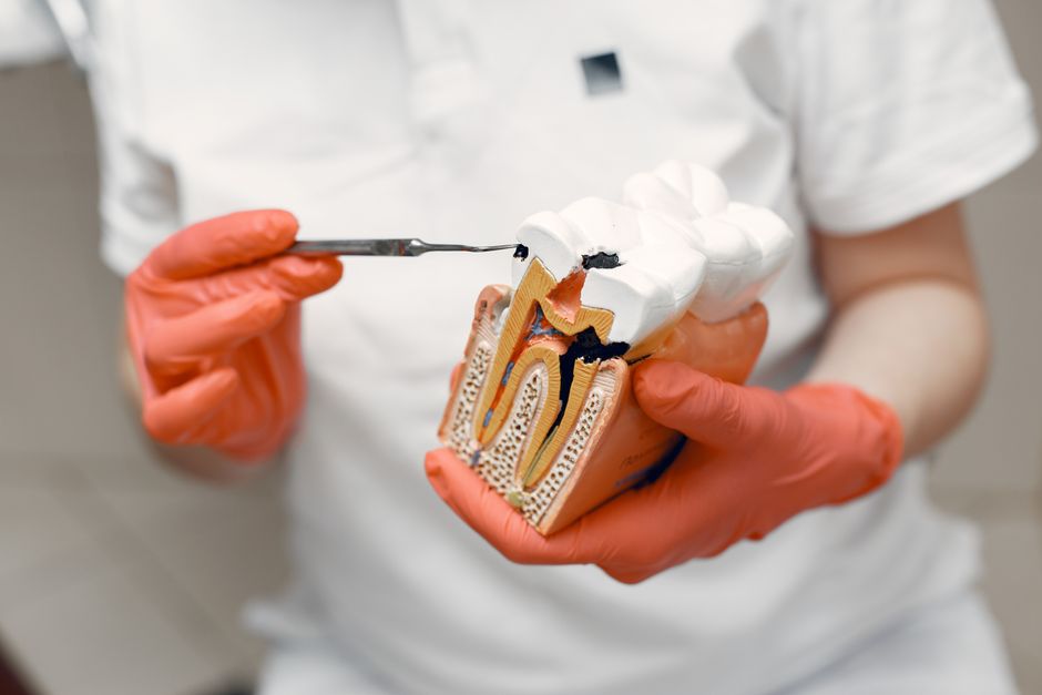
Material choices and biologic principles (what trainees should understand)
Augmentation materials can include autografts, allografts, xenografts, and alloplasts, often combined based on the desired remodeling rate and volume stability. Membranes and fixation methods support clot stability and space maintenance—two pillars of predictable regeneration. While published protocols vary, most educational frameworks emphasize:
1) Stable wound environment: minimize micromovement and contamination.
2) Space maintenance: prevent soft-tissue collapse into the defect.
3) Adequate blood supply: respect periosteal vascularity and avoid excessive thermal or mechanical trauma.
4) Tension-free closure: a core skill that often determines exposure risk.
Soft tissue and periodontal considerations: augmentation is not only “bone”
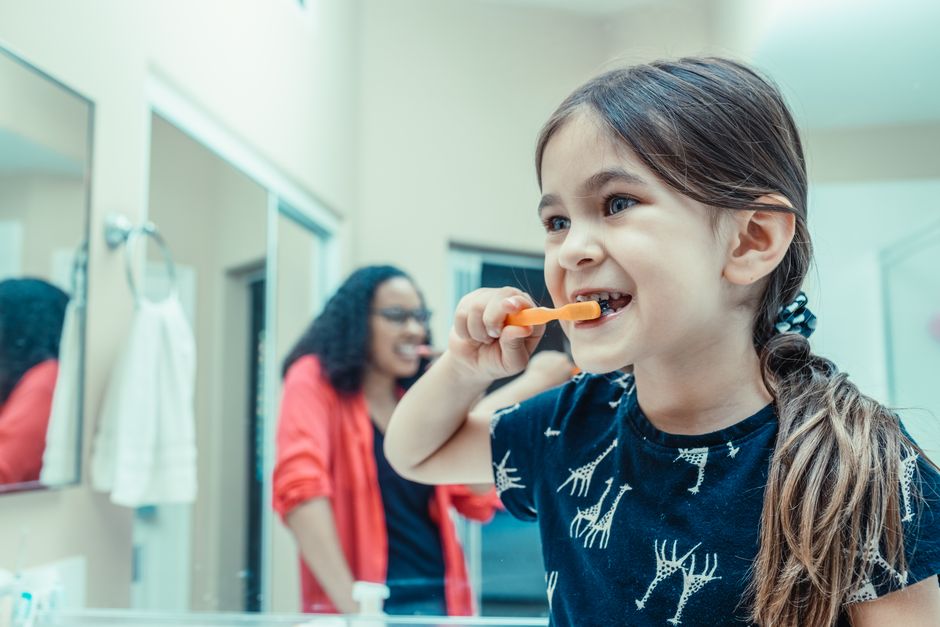
Long-term implant success depends on more than radiographic bone gain. Keratinized tissue width, mucosal thickness, and plaque control can influence comfort, cleansability, and peri-implant mucositis risk. Periodontal evaluation—both around remaining teeth and at future implant sites—should be part of the baseline workup. Patient education on home care is equally important; while implants differ from teeth, inflammation risk still relates strongly to biofilm.
For patients and clinicians interested in preventive habits, our educational content on evidence-informed ways to help prevent gum recession at home provides a practical lens on daily behaviors that can support soft-tissue stability—an area that matters before and after surgical site development.
Restorative-driven implant positioning: connecting augmentation to the final prosthesis
Augmentation should serve a restorative outcome: correct 3D implant placement, appropriate emergence, and a contour that supports hygiene. When bone is insufficient, clinicians may be forced to place implants too buccally/lingually or too apically, leading to esthetic compromise and difficult maintenance. This is why implant education increasingly integrates prosthodontics and digital planning early in the learning pathway.
In everyday practice, patients often compare implant therapy with purely cosmetic options. Understanding aesthetic case selection helps clinicians communicate ethically—what can be achieved with prosthetics alone versus what requires surgical site development. For a comparative perspective relevant to consultations, see Hollywood Smile vs zirconia crowns: clinical differences and case selection. While that topic focuses on restorative aesthetics, the decision-making mindset parallels implant planning: indications, limitations, and long-term maintenance should guide recommendations.
Managing adjacent problems: endodontics and restorative sequencing
Atrophic jaws often coexist with complex dental histories—failed endodontics, fractures, or periodontal breakdown. Treatment sequencing matters: teeth of questionable prognosis can influence augmentation timing and provisionalization strategies. When salvage is possible, high-quality endodontic treatment may preserve bone and delay or avoid implant needs; when extraction is indicated, atraumatic techniques and ridge preservation can reduce future atrophy.
Modern visualization tools can support precision in endodontics, which may indirectly support implant planning by preserving strategic teeth and alveolar housing. For clinicians upgrading their workflow, explore the dental operating microscope in modern endodontics—an example of how magnification and illumination integrate into contemporary multidisciplinary care.
Provisionalization, aesthetics, and patient expectations
In the anterior zone, patients may prioritize immediate aesthetics even when biology suggests staging. Provisional restorations (removable or fixed) should protect grafted sites, support soft-tissue shaping where appropriate, and remain cleanable. Overcontoured provisionals can compress tissues and complicate healing; under-supported tissues may collapse and limit aesthetic potential.
Aesthetic dentistry also intersects with implant planning when patients request veneers or smile enhancements around implant sites. The key educational point is that veneers, crowns, and implants should be planned together—shade mapping, incisal edge position, gingival zeniths, and occlusal scheme—so the final result looks cohesive.
If your practice includes veneer cases adjacent to implant sites, it is worth reviewing technique-sensitive pitfalls that can impact overall aesthetics and periodontal response. Our article on common mistakes in porcelain laminate veneers—and how to avoid them complements implant education by reinforcing conservative preparation principles, margin design considerations, and expectations management.
Complications and risk planning (educational, not prescriptive)
Even well-planned augmentation can face complications. Educational programs typically emphasize recognizing early warning signs and designing plans that reduce avoidable risks. Common challenges include membrane exposure, infection, graft mobility, insufficient volume gain, and patient-related factors such as smoking, uncontrolled systemic conditions, or inadequate plaque control.
Risk planning also includes anatomical considerations—sinus health, nerve proximity, and thin cortical plates—as well as logistic factors like follow-up compliance. For trainees, structured complication discussions (case reviews, debriefs, and decision trees) are often as valuable as the surgical steps themselves.
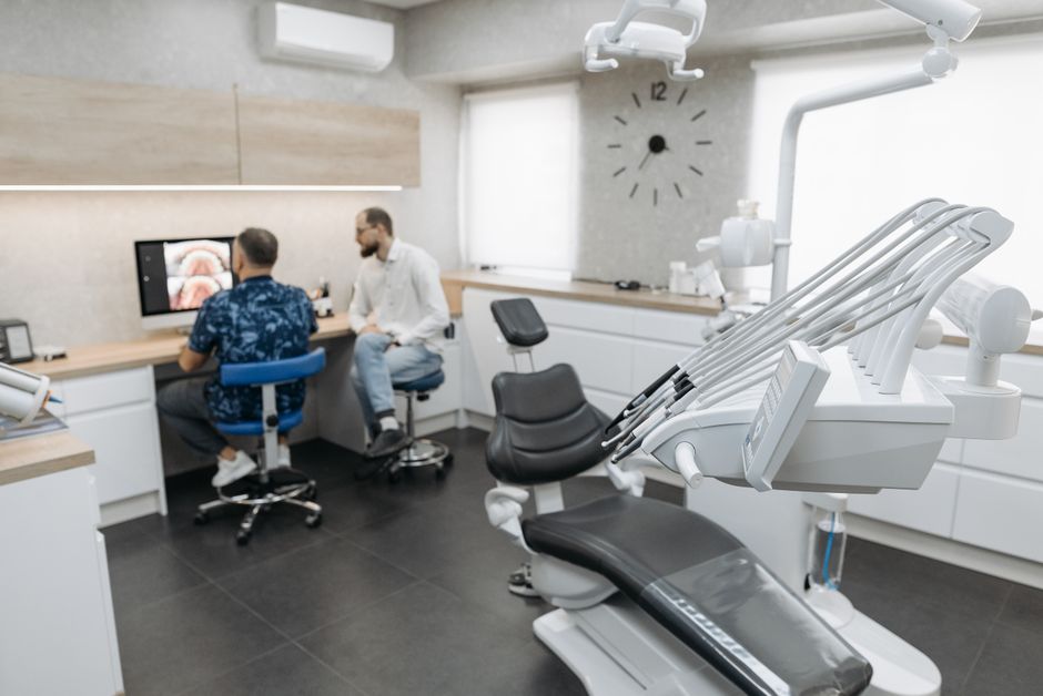
How Istanbul Dental Academy teaches atrophic jaw planning and augmentation
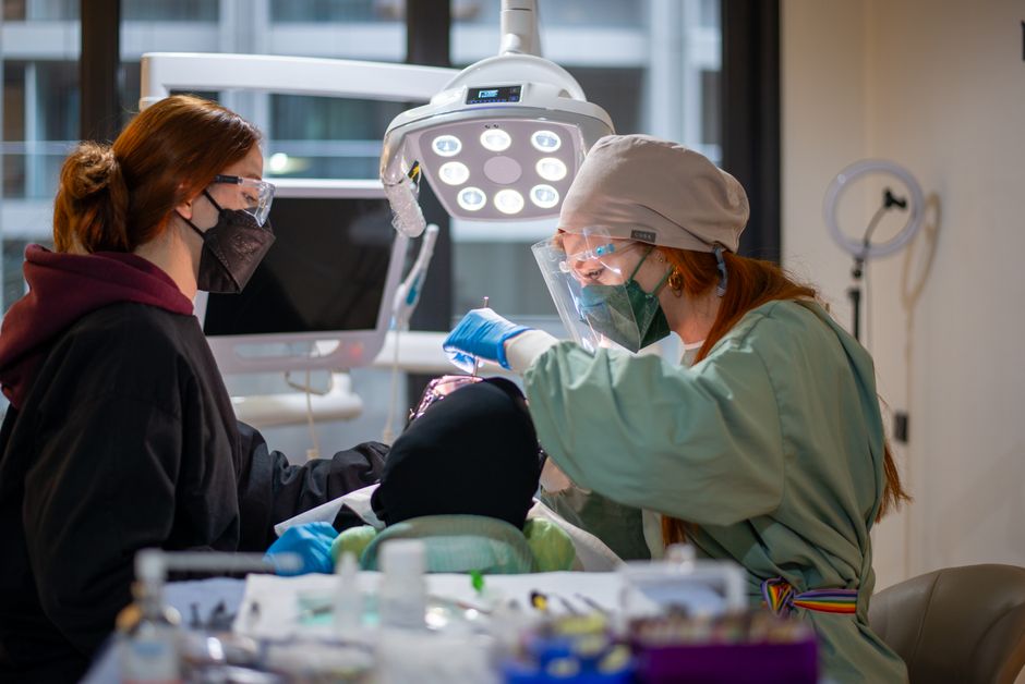
Because atrophic cases require integration of diagnostics, surgery, and prosthetics, clinicians often benefit from stepwise learning: foundational implant planning, flap and suturing skills, GBR principles, and then advanced modules such as sinus elevation, block grafting, and vertical augmentation concepts. At Istanbul Dental Academy, our educational focus is hands-on and case-based—participants learn how to read CBCT findings in context, translate restorative goals into surgical plans, and practice key maneuvers under guidance.
We also encourage clinicians to build supportive skills that elevate outcomes: digital workflows for planning and communication, dental photography for documentation and case presentation, and a maintenance mindset rooted in periodontal health. These competencies help clinicians manage expectations and track results responsibly.
Key takeaways
Atrophic jaws are not simply “less bone”—they represent a planning challenge where biology, anatomy, soft tissue, and prosthetic design converge. Bone augmentation before implant placement may be considered to enable ideal implant positioning and long-term maintainability, but technique selection should be individualized, risk-assessed, and aligned with the final restorative plan. This content is for educational purposes and is not a substitute for clinical training or patient-specific diagnosis. For clinicians seeking to deepen competence, structured continuing education and hands-on surgical training can help translate principles into predictable clinical execution.
Diğer Yazılar