BLOG
Could Severe Gum Pain Be Necrotizing Gingivitis? A Clinical Perspective
Blog Tarihi: 14/06/2026

When Severe Gum Pain Raises a Red Flag
Severe gingival pain is a complaint that can disrupt eating, sleeping, and oral hygiene routines—and it often prompts an urgent dental visit. While common gingivitis can cause discomfort, intense pain combined with specific clinical signs may suggest a more acute periodontal condition such as necrotizing gingivitis (often discussed under necrotizing ulcerative gingivitis, NUG).
For dental professionals, the value lies in rapid recognition, appropriate initial management, and thoughtful differential diagnosis. This content is for educational purposes and is not a substitute for individualized clinical judgment, diagnosis, or treatment planning.
What Is Necrotizing Gingivitis (NUG) and Why Can It Hurt So Much?
Necrotizing gingivitis is typically characterized by rapid onset gingival pain, necrosis of interdental papillae, and a distinctive clinical appearance. Pain severity can be disproportionate to the amount of visible plaque or calculus and may escalate quickly. Patients may describe a “burning” or “stabbing” sensation, sometimes accompanied by systemic symptoms such as malaise.
If you want a structured overview of hallmarks and clinical reasoning, see our dedicated guide: Necrotizing Ulcerative Gingivitis (NUG): Symptoms, Causes, and Clinical Approach.
Why pain can be severe
The pain associated with NUG is often linked to superficial tissue necrosis and acute inflammation, which can expose and irritate nerve endings. The resulting tenderness can discourage brushing and flossing, contributing to a vicious cycle of worsening local factors and microbial burden.
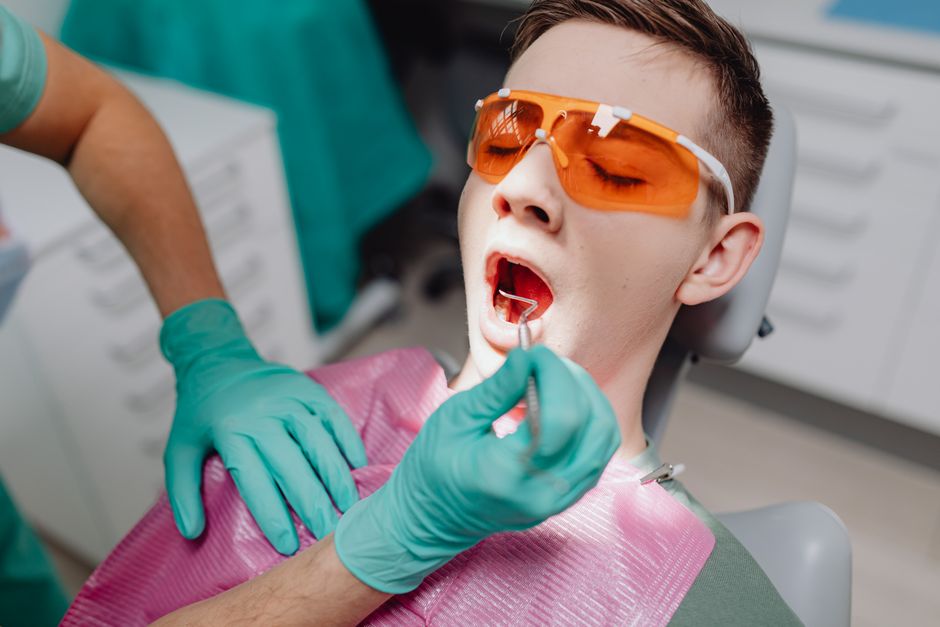
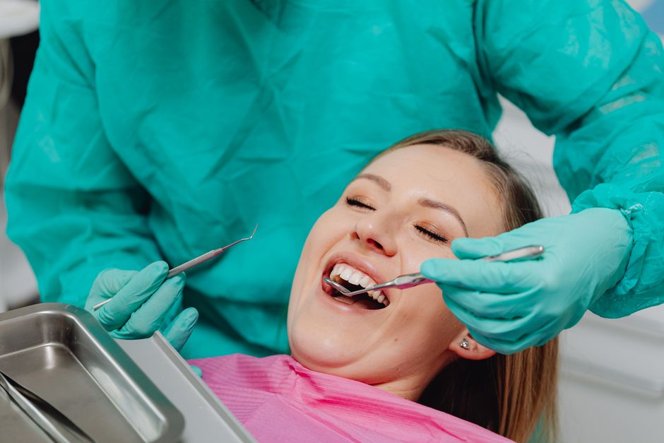
Clinical Signs: What to Look for Chairside
Although presentations vary, clinicians often consider NUG when severe pain occurs with several of the following features:
Key intraoral signs may include:

• “Punched-out” interdental papillae (necrosis at the tip)
• Spontaneous bleeding or bleeding on gentle probing
• Grayish pseudomembrane over affected gingiva
• Strong halitosis and metallic taste complaint
• Cratered papillae with erythema and edema in adjacent tissue
From a periodontal charting standpoint, careful documentation of bleeding, pain response, and lesion distribution helps establish baseline severity and supports follow-up comparisons. In acute pain cases, probing may be limited—clinical photography can help capture initial appearance for monitoring.
Risk Factors and Triggers: The “Perfect Storm” Scenario
NUG is frequently discussed as an opportunistic condition that can emerge when local biofilm challenges interact with systemic or behavioral stressors. While it is not appropriate to assume causality based on a single factor, clinicians commonly evaluate risk context as part of the diagnostic workup.
Commonly discussed risk contexts
• High psychosocial stress and sleep disruption
• Tobacco use
• Poor oral hygiene or abrupt decline in plaque control due to pain
• Immunocompromised states or systemic illness (case-dependent)
• Nutritional compromise or dehydration
• Recent respiratory infections
In Istanbul’s busy urban environment, it is not uncommon for patients—especially students and shift workers—to report stress, irregular meals, and skipped hygiene habits. Taking a brief, nonjudgmental history can clarify potential contributors and support patient education.
Differential Diagnosis: Not Every Painful Gum Lesion Is NUG
Severe gingival pain is a symptom, not a diagnosis. Conditions that may mimic aspects of NUG include acute herpetic gingivostomatitis, desquamative gingivitis (mucocutaneous disorders), aphthous-like ulcerations, aggressive periodontal infections, allergic/contact reactions, and trauma-related lesions. A careful review of lesion distribution, systemic symptoms, medical history, medication use, and the presence/absence of papillary necrosis is essential.
Additionally, odontogenic pain can radiate and be perceived as “gum pain.” Deep caries, irreversible pulpitis, cracked teeth, or acute apical pathology may present with localized tenderness near the gingiva—especially if swelling is present.
When endodontics enters the conversation
If the pain localizes near a specific tooth, responds to thermal stimuli, or is accompanied by percussion sensitivity, consider an endodontic assessment. In complex diagnostic situations, enhanced visualization can support clinician confidence. For an education-focused look at magnification and documentation, review: Dental Operating Microscope in Modern Endodontics: Better Vision, Better Outcomes.
Clinical Approach: Education-Focused Initial Management Considerations
Because NUG may present acutely and painfully, the first appointment often balances patient comfort with gentle debridement and risk assessment. While specific treatment decisions depend on the patient’s condition and clinician judgment, many protocols emphasize a staged approach: pain control, careful removal of necrotic debris and plaque in tolerable increments, and close follow-up.
Communication and patient cooperation
Patients in significant pain may fear brushing or eating. Chairside communication should normalize the experience while emphasizing that gentle plaque control and follow-up are critical. Short, clear instructions—supported by intraoral photos—can increase adherence during the acute phase.
Why follow-up matters
Even when symptoms improve quickly, relapse can occur if contributing factors persist. A structured re-evaluation allows reassessment of bleeding, tissue contour, and plaque control effectiveness, and supports transition into comprehensive periodontal care and long-term maintenance.
How Acute Gum Infections Affect Restorative and Esthetic Dentistry
From a restorative perspective, inflamed or necrotic gingiva can compromise impressions, isolation, bonding protocols, and margin placement. In esthetic cases—especially anterior smile makeovers—soft tissue stability is foundational. Attempting definitive esthetic procedures during active inflammation can lead to unpredictable outcomes, including margin discrepancies and patient dissatisfaction.
Smile design and veneers: the soft-tissue prerequisite
For clinicians planning laminate veneers, gingival health influences gingival zeniths, emergence profile, and the visual harmony of the smile. A modern workflow increasingly integrates digital planning, photography, and mock-ups—but periodontal stabilization remains a prerequisite. For a clinician-oriented overview, explore: Digital Dentistry for Laminate Veneer Planning: A Modern Smile Design Workflow.
Periodontal Health as the Foundation for Implant Dentistry
Although NUG primarily affects gingival tissues, the broader message for clinicians is that acute periodontal infections highlight the importance of infection control, patient risk profiling, and maintenance—principles that also apply to implant dentistry. Before implant placement, clinicians typically prioritize stable periodontal conditions and patient compliance with hygiene and recalls.
Digital implant workflows and diagnostic clarity
Digital dentistry can streamline implant planning and improve interdisciplinary communication. CBCT-based planning, intraoral scanning, and guided surgery workflows can support accuracy—especially in complex restorative-driven cases. To understand how digital tools integrate with implant treatment planning and workflow, read: The Role of CAD/CAM Technology in Modern Implant Dentistry.
When extraction and immediate implant placement are considered
Some patients present with pain and infection that ultimately requires extraction. While immediate implant placement can be an option in selected scenarios, decision-making is nuanced and depends on diagnosis, tissue condition, debridement feasibility, and overall risk assessment. For an educational clinical overview, see: Same-Day Tooth Extraction and Immediate Implant Placement: A Clinical Guide.
Training Takeaways for Dentists and Dental Students
NUG is a strong example of how periodontal diagnosis is not only about “seeing plaque,” but about reading tissue behavior, patient risk factors, and symptom intensity. For clinicians in continuing education, the case reinforces several practical competencies:
• Rapid recognition of acute periodontal signs and documentation standards
• Differential diagnosis between periodontal, endodontic, and mucosal pain sources
• Patient communication in urgent, pain-driven appointments
• Staged care planning: acute management, re-evaluation, and maintenance
• Interdisciplinary sequencing before esthetic, restorative, or implant procedures
At Istanbul Dental Academy, our hands-on courses emphasize clinical reasoning alongside technique—helping dentists integrate periodontal assessment into daily practice, improve photography and documentation habits, and plan interdisciplinary workflows that respect soft-tissue biology.
When Should a Patient Seek Urgent Dental Assessment?
From a public-facing standpoint, severe gum pain with bleeding, bad breath, or visible tissue changes warrants timely dental evaluation. Patients may also report fever, fatigue, or difficulty eating. While not every case is NUG, delaying assessment can increase discomfort and complicate care.
For clinicians, urgent appointments are an opportunity to stabilize symptoms, educate the patient, and create a stepwise plan that transitions from acute care to prevention and long-term periodontal maintenance.
Conclusion
Could severe gum pain be necrotizing gingivitis? In some cases, yes—especially when pain is intense and accompanied by papillary necrosis, bleeding, and halitosis. The key for dental professionals is to recognize the clinical pattern, rule out look-alike conditions, and manage the case in a staged, evidence-informed manner that supports long-term periodontal stability.
This content is for educational purposes only and does not provide definitive medical or treatment advice. Clinical decisions should be made by qualified dental professionals based on individual patient findings.
Diğer Yazılar


