BLOG
Hollywood Smile Before and After: Planning, Materials, and Clinical Considerations
Blog Tarihi: 25/06/2026
Hollywood Smile: what “before and after” really means
On social media, “Hollywood Smile before and after” often looks like a quick cosmetic switch. In daily practice, however, the “after” is typically the outcome of a sequence: diagnosis, risk control, aesthetic planning, minimally invasive preparation (when indicated), adhesive protocols, and maintenance. For dentists and dental students, the value of studying before-and-after cases is not only aesthetic—it is diagnostic. A strong result can be traced back to clinical decisions made long before the final photos are taken.
In Istanbul, where patients frequently seek comprehensive aesthetic rehabilitation during short visits, predictability and communication become even more critical. This is why continuing dental education increasingly focuses on integrated smile design workflows that combine restorative dentistry, prosthodontics, periodontology, and digital dentistry—skills emphasized in hands-on training at Istanbul Dental Academy. This content is for educational purposes and is not a substitute for individualized diagnosis or treatment planning.
Pre-treatment assessment: the “before” that determines the outcome
1) Chief complaint, expectations, and feasibility
The starting point is aligning expectations with biology and materials. “Whiter,” “straighter,” and “bigger” can conflict with periodontal limits, occlusal stability, and facial proportions. A structured interview helps clarify whether the patient is seeking a subtle enhancement, a high-value smile makeover, or a functional reconstruction disguised as an aesthetic request.
2) Periodontal status and soft-tissue architecture
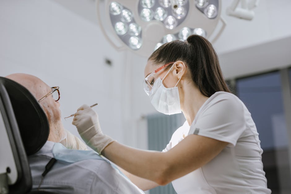
Gingival inflammation, bleeding on probing, and unstable margins can compromise impression accuracy, bonding, and long-term aesthetics. Before planning veneer margins or crown lengths, clinicians should identify periodontal phenotype, gingival zeniths, and smile line dynamics. Acute or severe gingival pain and necrotic presentations require urgent diagnostic consideration and stabilization before elective cosmetic work; for clinical context, see a clinical perspective on necrotizing gingivitis and severe gum pain.
In many “before” photos, subtle issues (edema, rolled margins, or uneven zeniths) later become obvious in the “after” when the teeth are brighter and the contrast increases. Periodontal re-evaluation and, when appropriate, coordination with a periodontist can prevent mismatched tissue levels and patient dissatisfaction.
3) Caries risk, erosion, and restorative baseline
Aesthetic cases often sit on a restorative foundation: old composites, leaking margins, erosion, or parafunction-related wear. The clinician’s job is to decide whether the case is primarily additive (bonding, veneers) or requires coverage and functional reorganization. Conservative options may include partial-coverage restorations; decision pathways are discussed in clinical decision-making on when to choose onlay vs overlay restorations.
4) Enamel quality and hypersensitivity considerations
Before preparing for veneers, evaluate enamel thickness, translucency, and developmental defects. Patients with enamel hypoplasia patterns or hereditary enamel conditions may present with sensitivity, altered bonding substrate, and atypical aesthetics. For a focused review relevant to restorative planning, consult clinical insights on amelogenesis imperfecta and tooth sensitivity.
5) Saliva, xerostomia, and adhesive predictability
Saliva is not only a comfort factor; it influences plaque control, mucosal health, and restorative longevity. Xerostomia can increase caries risk, contribute to soft-tissue irritation, and complicate bonding isolation. A “Hollywood Smile” plan should consider whether the patient’s oral environment supports long-term maintenance. For broader context, see Dry Mouth (Xerostomia): Causes, Risks, and Clinical Implications.
Smile design workflow: from digital plan to clinical reality
Facial and dento-labial analysis
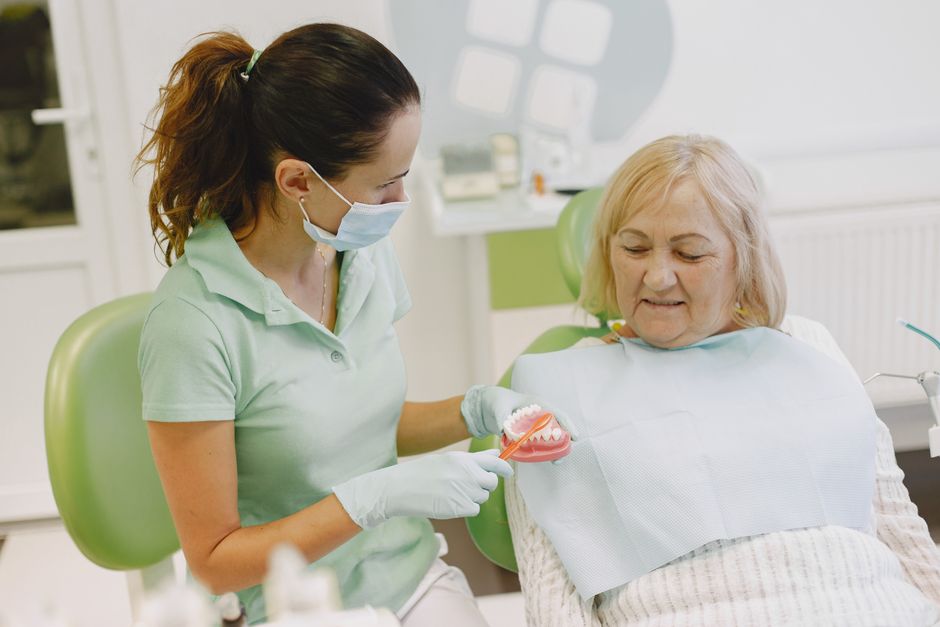
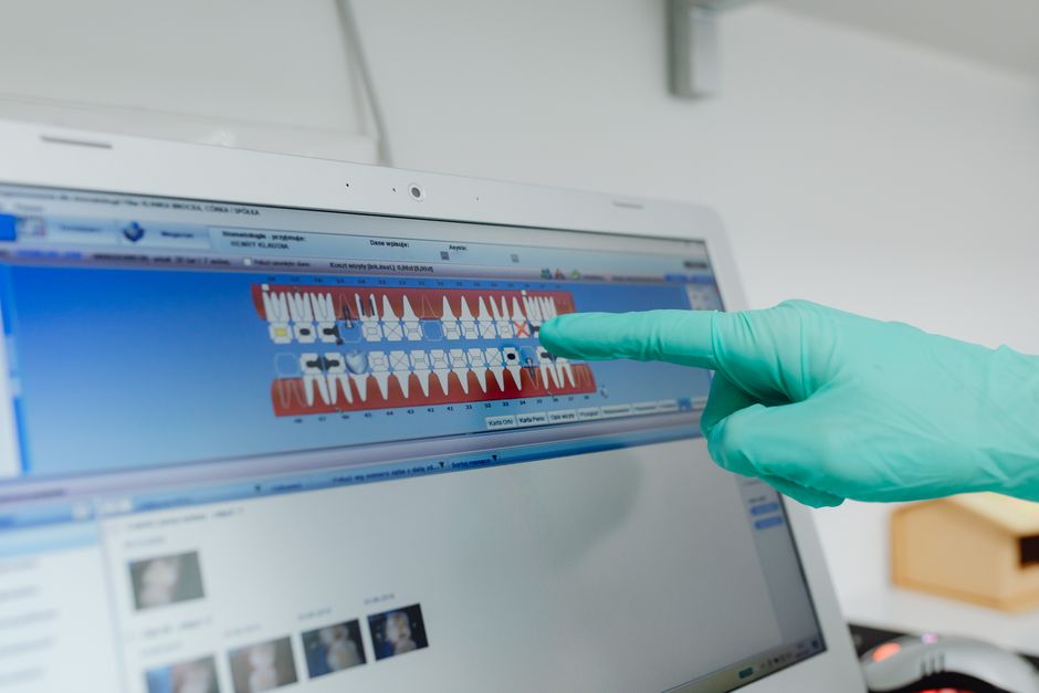
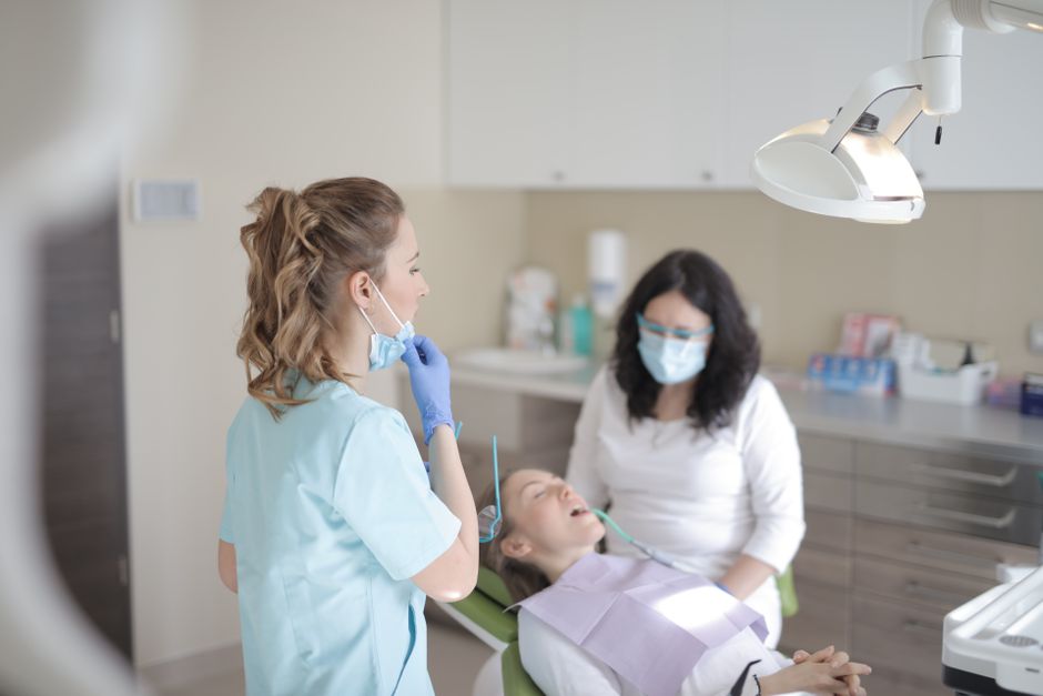
High-quality initial records are the bridge between “before” and “after.” Common elements include extraoral photos (rest and full smile), intraoral photos, videos to capture phonetics, and occlusal records. Facial midline, interpupillary line, smile arc, incisal display at rest, and lip mobility inform tooth length and gingival display decisions.
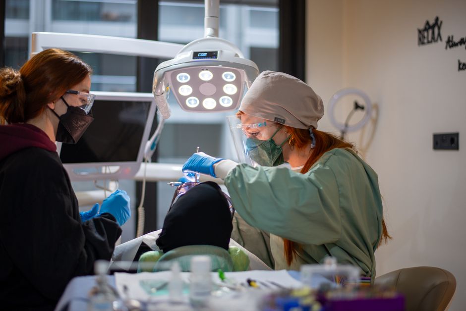
Dental photography becomes a clinical tool, not marketing. Standardized lighting, retraction, and calibration allow comparisons over time, improve lab communication, and support consent discussions. In education settings, structured photography protocols help dentists create replicable results rather than one-off successes.
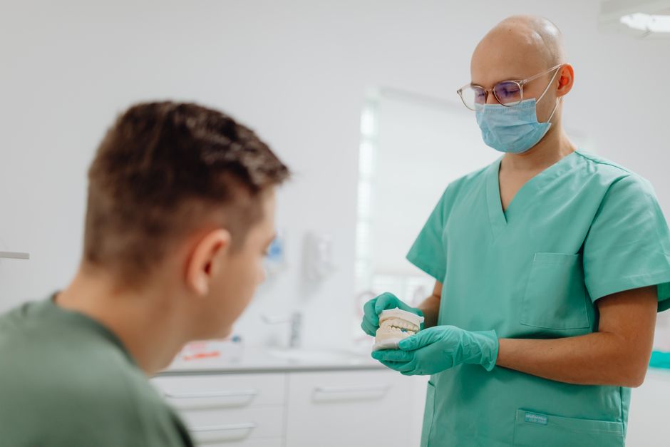
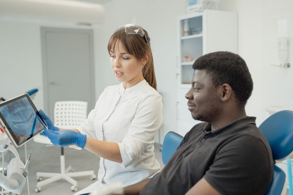
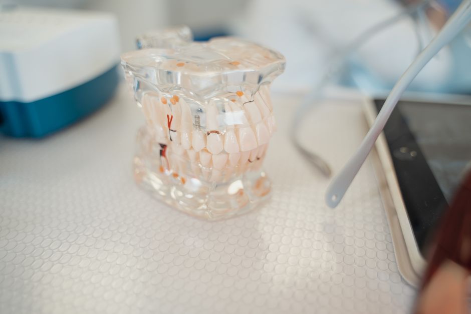
Digital Smile Design (DSD) and mock-ups
Digital planning can preview proportions and propose tooth shapes that harmonize with the face. Yet, the true “test drive” is often the intraoral mock-up (direct composite or bis-acryl based on a wax-up). Mock-ups allow the patient and clinician to evaluate phonetics (“F” and “V” sounds), lip support, and perceived tooth dominance before enamel is touched.
In short-stay cases common in Istanbul, a validated mock-up reduces remakes and chair-time stress. For clinicians, learning a repeatable DSD-to-mock-up workflow is a practical skill set developed in hands-on modules at Istanbul Dental Academy, where restorative planning is taught alongside preparation design and adhesive execution.
Common treatment components in “Hollywood Smile” cases
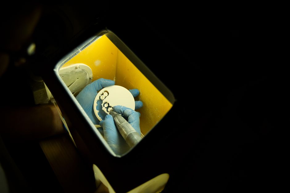
Porcelain laminate veneers: indications and limitations
Porcelain laminate veneers are often associated with the Hollywood Smile because they can deliver high esthetics with conservative preparation when case selection is appropriate. Ideal indications may include color modification, mild shape correction, closure of small diastemas, and surface texture enhancement—provided occlusion and parafunction are managed.
However, veneers are technique-sensitive: margin placement, isolation, and bonding protocols can determine longevity. For clinicians refining adhesive steps, review critical points in porcelain laminate cementation, which highlights areas where small errors can lead to sensitivity, marginal discoloration, or debonding.
Restorative dentistry: additive vs subtractive planning
Many “after” smiles are the result of additive dentistry—building the ideal contours with minimal reduction. When tooth volume must be reduced (e.g., protrusion, severe discoloration requiring masking, or alignment compromises), clinicians should document why reduction is necessary and how much is planned. Depth-cut guides, reduction keys, and preoperative wax-ups increase control and reduce over-preparation.
Periodontal and surgical refinements
Uneven gingival margins, altered passive eruption, or asymmetrical zeniths can dominate the final appearance. Depending on diagnosis, crown lengthening or soft-tissue recontouring may be considered. Surgical timing matters: rushing restorative finalization before tissue stabilization may compromise margin placement and “after” photo symmetry. Coordinated planning between prosthodontics and periodontology is a hallmark of predictable aesthetic dentistry.
Implant dentistry and prosthodontics in aesthetic zones
Some smile makeovers extend beyond veneers—missing teeth, failing crowns, or hopeless roots may require implant-based solutions or fixed prosthodontics. In the aesthetic zone, emergence profile, papilla management, and provisionalization strategy are often more important than the implant fixture itself. Photographic documentation and a staged approach can help patients understand why an implant case may not deliver an “instant” transformation.
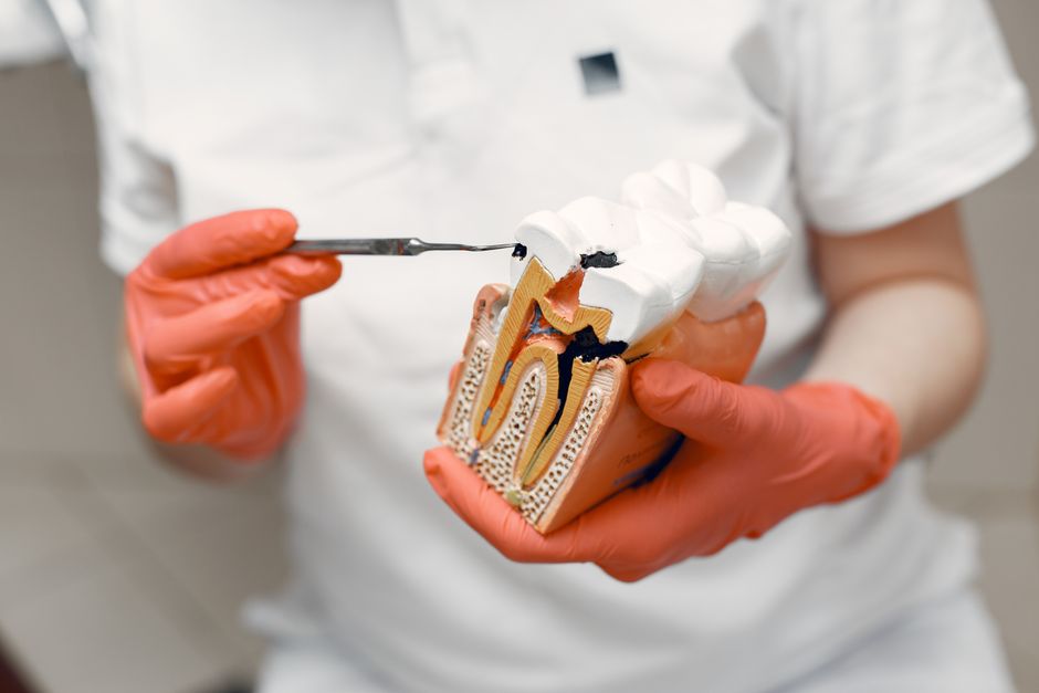
Endodontics: the hidden step behind aesthetic outcomes
Discolored teeth, trauma history, or deep restorations may require endodontic assessment prior to cosmetic procedures. A stable endodontic baseline helps reduce postoperative complications and supports restorative choices. Even when the patient asks only for a whiter smile, clinicians should verify pulpal status and periapical health as part of comprehensive care.
Before-and-after pitfalls: what can go wrong (and how to reduce risk)
Over-whitening and value mismatch
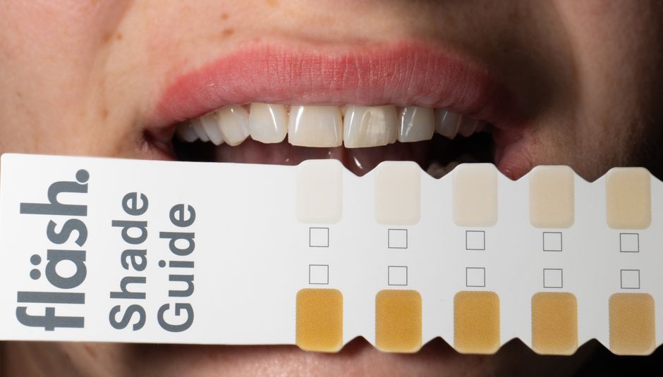
Very high-value shades can flatten tooth anatomy and challenge lip/skin harmony, especially under natural daylight. Texture, translucency, and incisal effects influence whether the smile looks “premium” rather than artificial. Shade selection should be discussed with the patient using calibrated photos and, where possible, try-in pastes for veneers.
Ignoring occlusion and parafunction
Bruxism and unstable occlusion can lead to chipping, debonding, and fractured ceramics. Before-and-after galleries rarely show nightguard compliance, but maintenance planning is often what protects the “after.” In education, occlusal assessment is integrated into aesthetic workflows to help clinicians design restorations that look good and function well.
Soft-tissue inflammation at delivery
Inflamed gingiva at cementation or delivery can distort margins and compromise bonding. Tissue management (retraction strategy, hemostasis control, and provisional contouring) should be treated as core restorative skills rather than optional extras. When inflammation is present, clinicians should consider postponing definitive steps until the etiology is addressed and tissues are stable.
How to document “before and after” ethically and professionally
Photography standards for clinical communication
Consistent framing, retractors, mirrors, and color calibration improve both diagnosis and credibility. For professional education and patient communication, include retracted frontal, right/left buccal, occlusal views, and face shots at rest and smile. Using the same settings and background makes the comparison meaningful rather than marketing-driven.
Consent, transparency, and realistic messaging
Ethical before-and-after documentation should disclose the type of procedures performed and avoid implying guaranteed outcomes. Small differences in lighting and lip posture can exaggerate results. A transparent workflow supports patient trust and reduces misunderstandings—especially important for high-visibility cosmetic cases.
Learning the workflow: why hands-on training matters
Hollywood Smile cases are rarely one-skill cases. They require integrated competence in diagnosis, smile design, preparation principles, soft-tissue management, adhesive dentistry, and finishing/polishing. While reading and watching cases can improve understanding, tactile experience—preparing, bonding, adjusting occlusion, and troubleshooting—is often what converts knowledge into predictable outcomes.
Istanbul Dental Academy structures continuing dental education around practical workflows: digital planning to mock-up, minimally invasive preparation strategies, and cementation protocols supported by clinical photography and case discussion. For dentists aiming to deliver high-end aesthetic outcomes responsibly, a course pathway that combines restorative dentistry, prosthodontics, and digital dentistry can be a clinically relevant next step.
Key takeaways for clinicians
“Before and after” is not a single moment—it is the visual summary of diagnosis, sequencing, and execution. Strong outcomes typically come from (1) controlling periodontal and caries risk, (2) validating aesthetics with mock-ups, (3) choosing materials based on substrate and function, and (4) applying meticulous adhesive and tissue-management protocols. This content is for educational purposes; definitive decisions should be made through individualized clinical evaluation, diagnostics, and informed consent.
Diğer Yazılar