BLOG
Why Imaging Technologies Matter in Modern Root Canal Treatment
Blog Tarihi: 26/06/2026
Imaging as the “Navigation System” of Endodontics
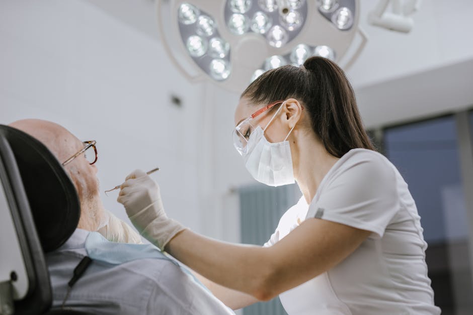
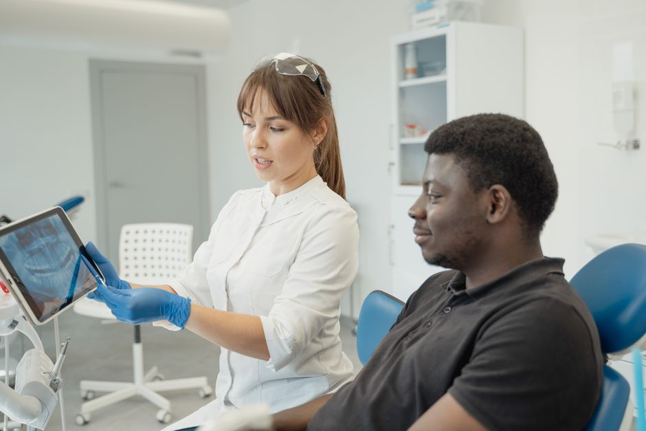
Root canal treatment is often described as a procedure performed “blind.” In reality, modern endodontics is guided by imaging—before, during, and after treatment. High-quality diagnostic images help clinicians identify apical pathosis, locate missed anatomy, assess restorability, and evaluate complications such as perforations or separated instruments. In daily practice, imaging is not only a documentation tool; it is a clinical decision-making instrument that directly influences case selection, treatment planning, and follow-up protocols.
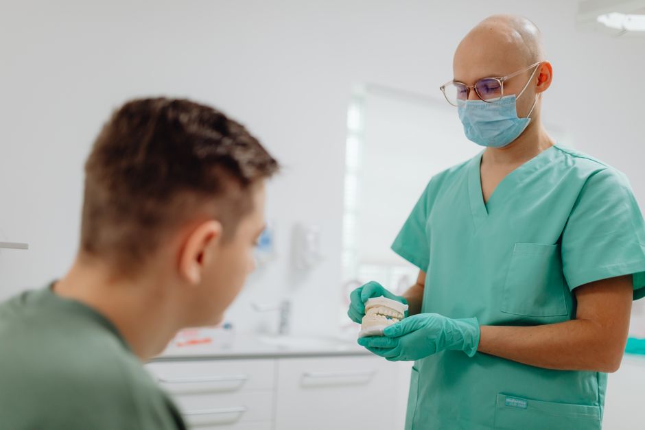
For dentists and dental students seeking continuing education, understanding how to interpret radiographs and when to choose advanced imaging can be as important as mastering instrumentation. At Istanbul Dental Academy, imaging literacy is integrated into hands-on learning, because predictable outcomes in endodontics, restorative dentistry, prosthodontics, and even implant dentistry start with accurate diagnosis and proper workflow planning. This content is for educational purposes and does not replace individualized clinical judgment or patient-specific assessment.
Core Imaging Modalities in Root Canal Treatment
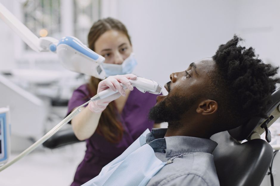
Periapical Radiography: Still the Workhorse
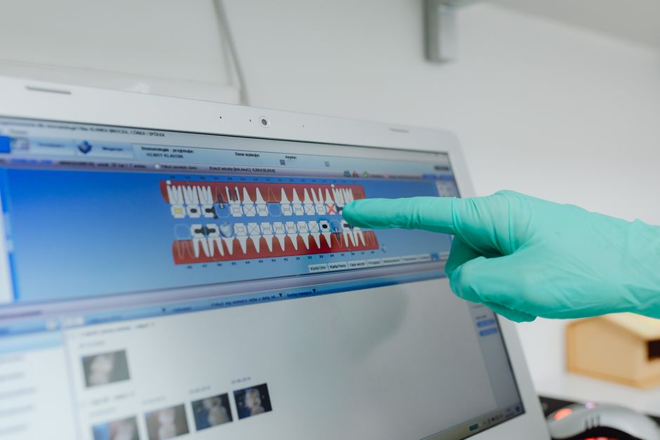
Despite rapid digital advances, periapical radiographs remain the foundational imaging method in endodontics. With careful angulation and exposure control, they can reveal caries depth, periodontal support, periapical radiolucencies, and approximate canal anatomy. Digital sensors and phosphor plates have improved speed, image enhancement options, and patient record management.
However, clinicians must remain aware of limitations: periapicals are two-dimensional representations of three-dimensional structures. Superimposition can conceal extra canals, root fractures, or the true extent of lesions. This is why multiple angulated images (when clinically appropriate) and a systematic interpretation approach are essential.
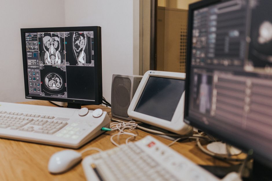
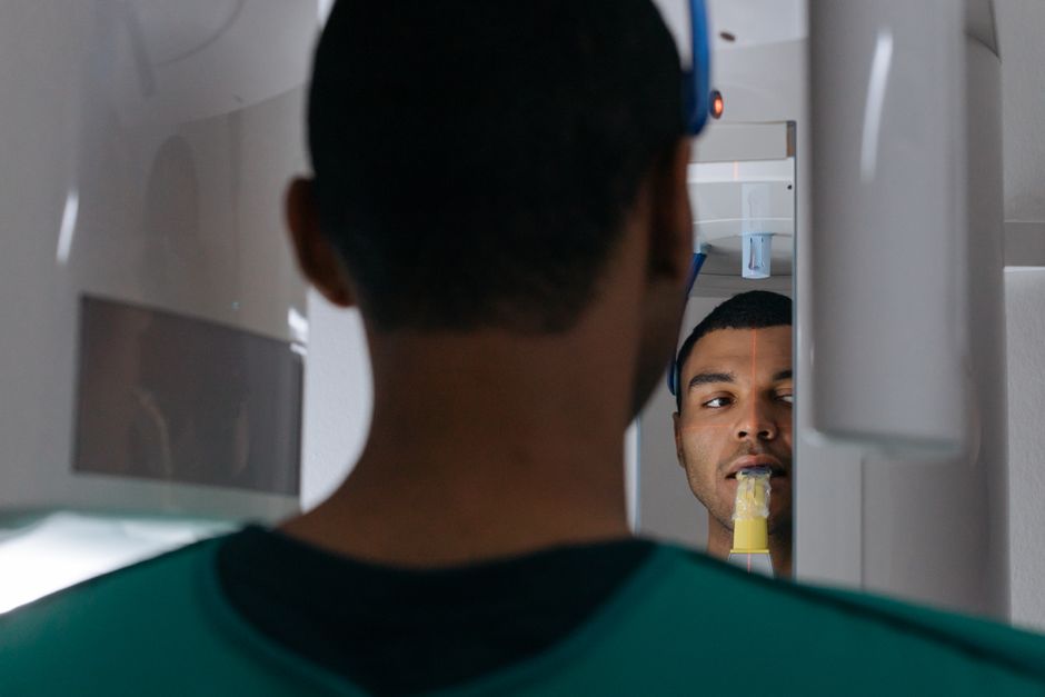
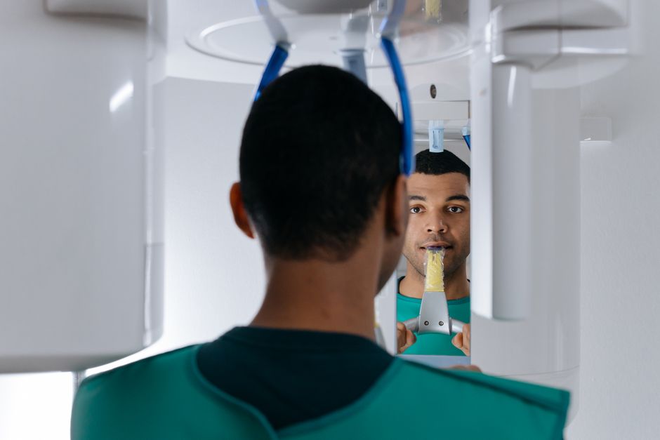
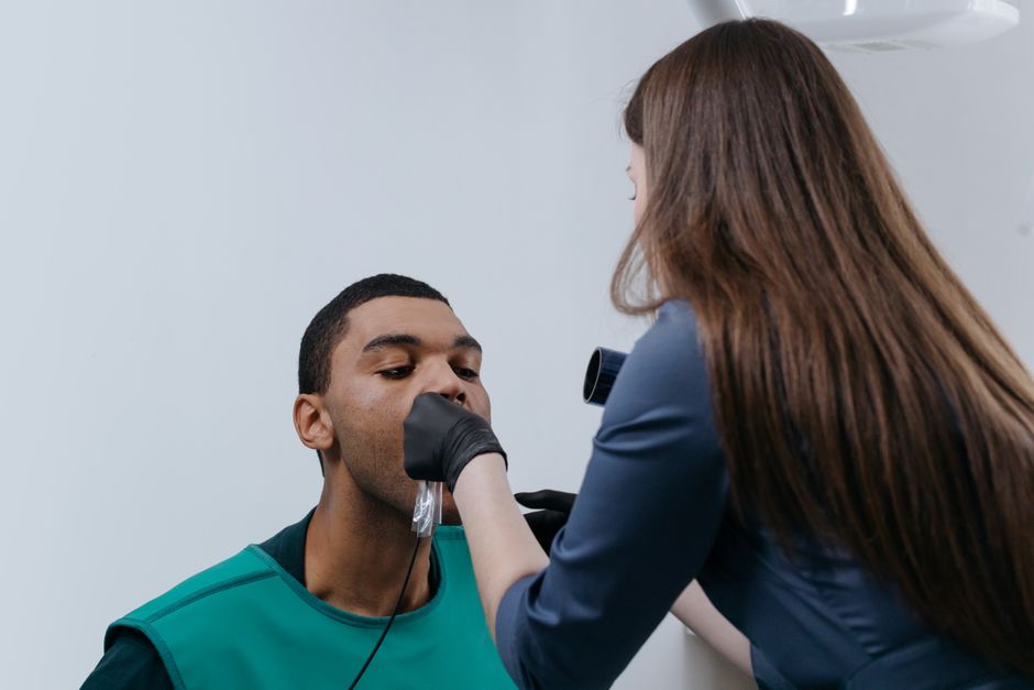
CBCT (Cone Beam CT): When 3D Changes the Diagnosis
CBCT can provide three-dimensional visualization of root canal anatomy and surrounding structures, often clarifying findings that are ambiguous on periapical radiographs. In endodontics, CBCT may be considered in scenarios such as suspected vertical root fracture, complex retreatment, resorptive defects, unusual anatomy, or pre-surgical planning. It can also help evaluate proximity to anatomical landmarks (e.g., mandibular canal, maxillary sinus) when planning endodontic microsurgery or assessing risks.
CBCT is not a “routine add-on.” Appropriate case selection, radiation awareness, and correct field-of-view choice are central to responsible use. The learning curve is also real: artifacts from metallic restorations, motion, or beam hardening can mislead interpretation. For this reason, structured education on indications, interpretation, and reporting is crucial for clinicians who want to incorporate CBCT into their endodontic workflow.
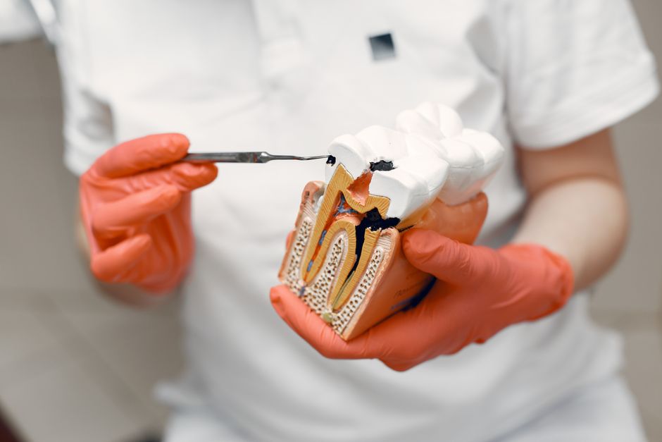
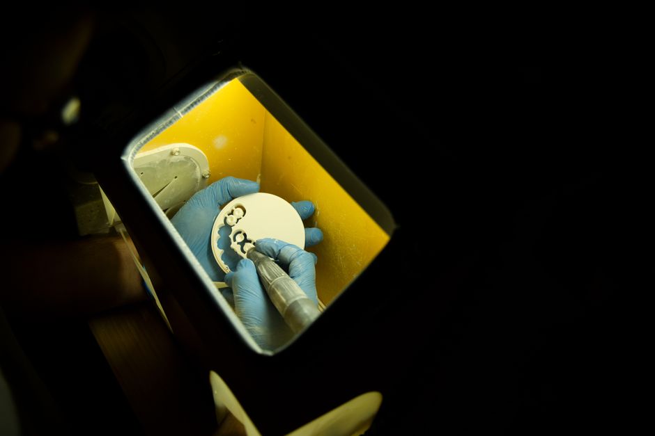
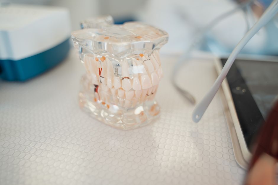
Magnification and Illumination: Imaging Beyond Radiographs
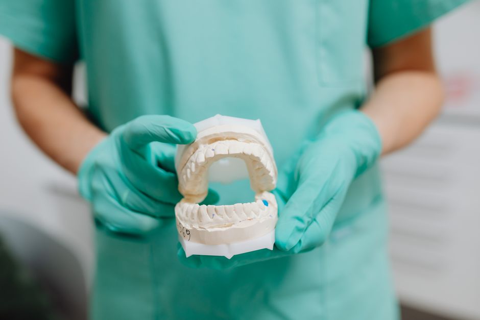
While magnification is not “imaging” in the radiographic sense, loupes and dental operating microscopes provide a different form of visual information—micro-anatomical visualization. Enhanced illumination and magnification can improve detection of calcified canals, cracks, isthmuses, and pulp chamber landmarks. This is especially relevant in retreatment cases, where missed anatomy and restorative obstructions are common.
In hands-on settings, clinicians often report that microscope training changes not only what they see, but how they plan access design, how conservatively they cut dentin, and how confidently they manage procedural challenges.
Imaging Across the Endodontic Workflow
1) Preoperative Diagnosis and Case Selection
Preoperative imaging supports differential diagnosis: pulpal inflammation versus necrosis, odontogenic pain versus referred pain, and endodontic lesions versus periodontal pathology. In multidisciplinary practice, this diagnostic clarity becomes even more important when patients present with additional concerns such as occlusal discomfort or joint-related symptoms—areas where broader clinical reasoning matters. For clinicians who want to expand diagnostic skills beyond the tooth, Istanbul Dental Academy’s evidence-informed resources—such as Clinical Diagnostic Approach to TMD Patients: An Evidence-Informed Guide for Dentists—highlight how structured evaluation can reduce misdiagnosis and improve communication across specialties.
From an endodontic standpoint, imaging also helps determine restorability: remaining tooth structure, ferrule potential, and the feasibility of coronal seal. A well-executed root canal cannot compensate for a non-restorable tooth.
2) Access Cavity Planning: Preserving Tooth Structure
Access is where imaging meets minimally invasive dentistry. Radiographs—and in complex cases, CBCT—help estimate pulp chamber depth, canal orientation, and the presence of restorative obstacles. Conservative access designs aim to preserve pericervical dentin and reduce fracture risk while maintaining adequate straight-line access. The balance between conservation and visibility is a clinical skill that improves through feedback-based training.
This principle links endodontics directly to restorative outcomes. When anterior teeth are involved, the endodontic access design and subsequent build-up influence esthetics, translucency, and final restoration selection. If you are interested in how trauma and structural loss are managed in the esthetic zone, see Contemporary Approaches to Restoring Fractured Anterior Teeth for a restorative perspective that often intersects with endodontic planning.
3) Working Length, Shaping, and Obturation Control
Working length determination is typically supported by electronic apex locators, with radiographs used to confirm file position and overall canal trajectory. Imaging helps identify potential ledges, transportation, and apical curvature—factors that inform instrument choice and motion strategy. During obturation, a final radiograph documents length, density, taper, and any voids that may compromise sealing.
Quality control becomes more reliable when clinicians adopt a consistent imaging protocol: standardized positioning, consistent angulation when comparing follow-ups, and careful documentation of baseline status. Digital workflows also allow easier archiving and case review—useful for both clinical auditing and educational portfolios.

4) Postoperative Assessment and Follow-Up
Healing in endodontics is evaluated over time. Imaging assists in monitoring reduction of periapical radiolucency, lamina dura continuity, and the absence of new lesions. Yet radiographic healing may lag behind clinical improvement. A systematic recall strategy supports evidence-informed care and helps clinicians communicate realistic expectations to patients.
When Endodontic Imaging Connects to Restorative and Prosthodontic Decisions
Posterior Teeth: Occlusion, Coverage, and Longevity
In posterior teeth, imaging findings influence whether a tooth should receive a direct composite, an onlay, or full-coverage restoration after endodontic treatment. Remaining cusp thickness, crack lines, and restoration margins are all part of the decision. For clinicians refining their restorative workflows alongside endodontic protocols, Direct vs Indirect Posterior Restorations: Clinical Decision-Making and Modern Workflows provides a practical framework that complements endodontic case planning.
Anterior Esthetics: Smile Design Planning Starts with Diagnostic Records
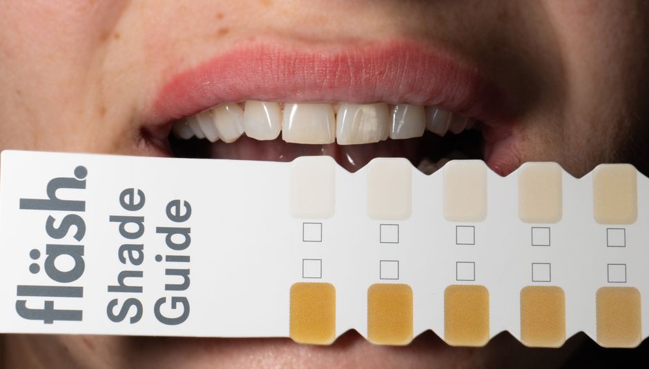
Imaging is also relevant in cosmetic workflows. Before porcelain laminate veneers or broader smile design cases, clinicians often need to confirm pulpal status, assess previous endodontic treatment quality, and rule out periapical issues. A tooth with undiagnosed apical pathology can undermine esthetic outcomes and patient satisfaction.
In patient communication, timelines matter. Diagnostic imaging supports realistic scheduling—especially when endodontic stabilization must occur before elective cosmetic steps. For a broader look at planning and longevity expectations, explore How Long Does Smile Design Take—and How Long Does It Last?, which highlights how sequencing and case complexity influence treatment duration.
Imaging in Complicated Cases: Retreatment, Resorption, and Surgery
Retreatment and Missed Anatomy
Retreatment cases are frequently imaging-driven. Identifying missed canals, untreated anatomy, or apical transportation may require careful periapical series and, in selected cases, CBCT. The clinician’s goal is not simply to “redo” the root canal, but to understand why failure occurred and whether correction is predictable.
Resorption and Suspected Root Fracture
Internal and external resorption can be difficult to characterize in 2D images. CBCT may assist in determining lesion location, extent, and proximity to the canal, which informs prognosis and treatment options. Similarly, suspected vertical root fractures can be diagnostically challenging; while CBCT may provide additional information, interpretation must account for artifacts and clinical signs.
Endodontic Microsurgery Planning
When non-surgical retreatment is not feasible or has a limited prognosis, imaging supports surgical planning: root apex location, cortical thickness, lesion boundaries, and adjacent anatomical structures. CBCT may enhance risk assessment and help determine flap design and access route. As with all advanced procedures, competence depends on training, supervision, and case selection.
Periodontal Considerations: Differentiating Endo-Perio Patterns
Imaging plays a key role in distinguishing primary endodontic lesions from periodontal disease patterns, particularly when probing depths and radiographic bone loss suggest combined involvement. A comprehensive periodontal evaluation and medical history review are essential, because inflammation and infection may coexist across tissues.
Clinicians should also recognize that acute gingival infections can complicate examination and patient comfort. For a periodontal-focused clinical overview, Managing Necrotizing Ulcerative Gingivitis (NUG): Clinical Steps and Prevention offers educational insights into assessment and prevention strategies that may be relevant when differential diagnosis is challenging.
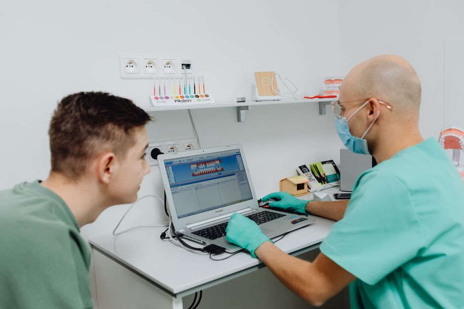
Digital Dentistry and Documentation: From Diagnosis to Education
Standardized Imaging Protocols Improve Consistency
Digital dentistry is not limited to CAD/CAM restorations—diagnostic imaging is a cornerstone. Standardized periapical techniques, clear labeling, consistent exposure settings, and organized record systems help clinicians track outcomes and share cases for peer discussion. For dental students and early-career dentists, building this habit early improves both clinical confidence and medico-legal documentation quality.
Dental Photography Complements Radiographic Imaging
In endodontics, photography can support documentation of crack lines, access cavity design, canal location, isolation quality, and restorative steps. Combined with radiographic records, clinical photos can strengthen patient communication and enhance learning—especially when reviewing cases in study groups or continuing education settings.
How Istanbul Dental Academy Approaches Imaging in Hands-On Learning
At Istanbul Dental Academy, imaging is treated as a clinical competency, not just a theoretical topic. In hands-on endodontic and restorative education, participants learn how imaging informs decision-making: selecting appropriate cases, planning access, anticipating anatomic challenges, and evaluating outcomes. Courses that integrate digital workflows, magnification, and documentation aim to support dentists who want to practice more predictably and communicate more clearly with patients and interdisciplinary colleagues.
Because imaging technologies and guidelines evolve, continuing dental education helps clinicians remain current—particularly when incorporating CBCT interpretation, microscope-based workflows, and digital record standards into everyday practice.
Key Takeaways
Imaging technologies are essential to modern root canal treatment: they guide diagnosis, reduce uncertainty during treatment, and support long-term follow-up. Periapical radiography remains indispensable, CBCT can be transformative in selected complex cases, and magnification improves visualization of micro-anatomy that radiographs cannot show. When integrated thoughtfully, imaging strengthens the connection between endodontic success and restorative longevity—supporting better outcomes in posterior rehabilitation, anterior esthetics, and comprehensive dental care.
This content is for educational purposes only and is not a substitute for professional training, local regulations, or individualized patient assessment. For clinicians looking to deepen practical skills, structured hands-on education can help translate imaging knowledge into safer, more predictable clinical workflows.
Diğer Yazılar