BLOG
Bruxism and TMJ Disorders: Clinical Links, Diagnosis, and Education
Blog Tarihi: 25/06/2026

Understanding the Bruxism–TMJ Connection in Daily Practice
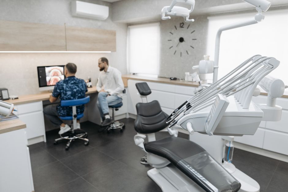
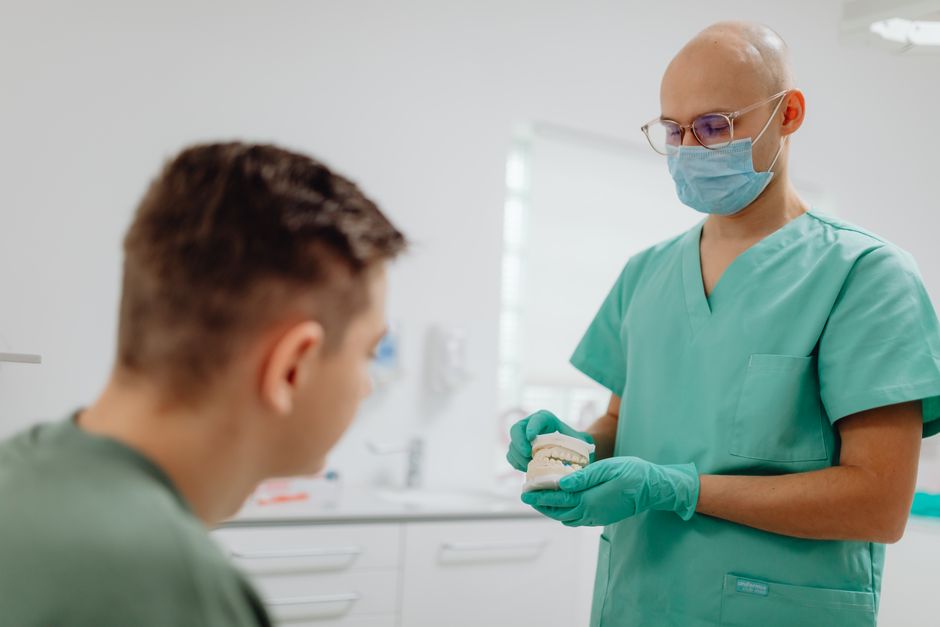
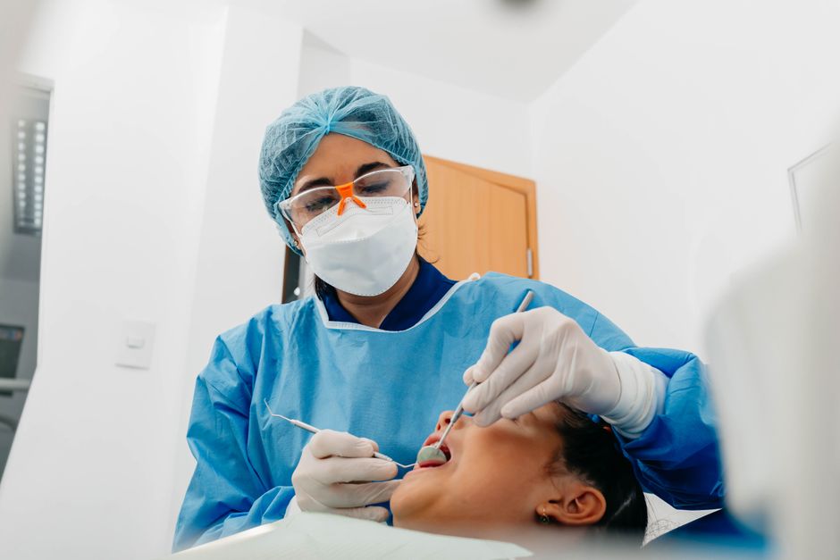
Bruxism—typically described as repetitive jaw-muscle activity such as clenching or grinding—frequently appears in the same clinical conversations as temporomandibular joint (TMJ) disorders (often grouped under the broader term temporomandibular disorders, TMD). In Istanbul clinics, as in many global settings, patients rarely present saying “I have bruxism” or “I have a TMJ disorder.” Instead, they report headaches, jaw fatigue, clicking, fractured restorations, morning soreness, or a sense that their bite “feels off.”
The key clinical challenge is that bruxism and TMD can be associated, but not in a simple cause-and-effect way. Some bruxers have no TMJ symptoms; some TMD patients do not grind; and many symptoms are influenced by stress, sleep quality, airway factors, parafunction, and occlusion. This article is for educational purposes and aims to help dental professionals structure a practical, evidence-informed approach—while highlighting why hands-on continuing education is valuable when integrating occlusal concepts into restorative dentistry, prosthodontics, and smile design.
Definitions That Matter: Bruxism vs. TMD
Bruxism: Awake vs. Sleep Presentations
Contemporary classifications distinguish between awake bruxism (often sustained clenching during daytime concentration or stress) and sleep bruxism (episodic jaw-muscle activity during sleep). The patient’s awareness, triggers, and risk profile can differ between the two. Importantly, bruxism is not automatically a disorder; it may be a behavior or a risk factor that becomes clinically relevant when it leads to tissue damage, pain, or functional limitation.
TMD: A Group of Conditions, Not a Single Diagnosis
TMD encompasses multiple diagnoses, including myalgia (muscle-related pain), arthralgia (joint pain), disc displacement with or without reduction (clicking or locking patterns), degenerative joint disease, and more. A patient with joint sounds but no pain or limitation may not require the same approach as a patient with significant jaw pain and restricted opening. Precise terminology improves communication, records, referrals, and treatment planning.
How Bruxism and TMD May Be Linked
Shared Risk Factors and Overlapping Symptoms
Bruxism and TMD often share risk factors such as psychosocial stress, anxiety, sleep disturbances, and certain behavioral patterns. Symptom overlap can be substantial: both may present with morning jaw fatigue, temple pain, or tooth sensitivity. Because of this overlap, clinicians sometimes over-attribute TMD pain to bruxism alone, or assume that any wear facets equal a TMJ disorder.
Biomechanical Load: Muscles, Joint, and Teeth
Excessive or frequent jaw-muscle activity can increase load on the masticatory system. In some patients, this may correlate with muscle pain (myofascial pain), tension-type headaches, or tenderness on palpation. In others, joint structures may be involved, especially if there is pre-existing disc or degenerative change. Yet, many individuals tolerate high load without pain—suggesting individual susceptibility, adaptation capacity, and central pain processing all play roles.

Restorations Under Stress: Clinical Red Flags
From a restorative perspective, bruxism may manifest as repeated ceramic chipping, debonding, crack lines, or accelerated wear of composite and enamel. These restorative failures can coexist with TMD symptoms, but they also occur in patients with no TMJ complaints. When treatment planning veneers, full-mouth rehabilitation, or implant restorations, documenting parafunctional risk and discussing protective strategies is a professional standard.
Clinical Assessment: A Stepwise Workflow
History: Ask Targeted Questions
Efficient screening questions can reveal patterns: When is pain worse—morning or evening? Any jaw locking? Headaches? Ear-related symptoms? Noise or clicking? History of trauma? Stress level? Sleep quality? Medication changes? Reports from partners about grinding? Prior splint therapy? The goal is to identify the dominant complaint (pain, function, or damage) and categorize likely contributors.
Extraoral and Intraoral Examination
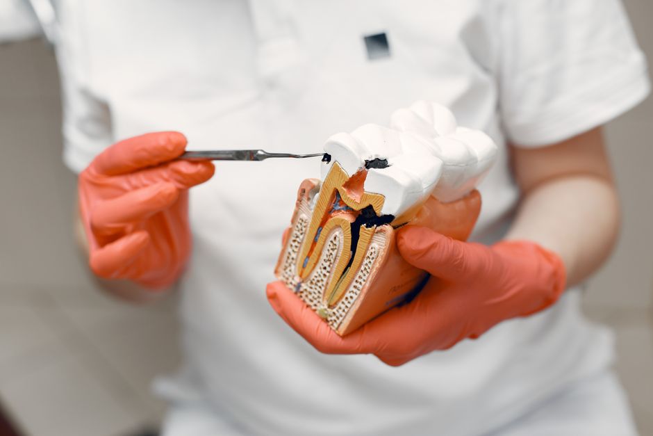
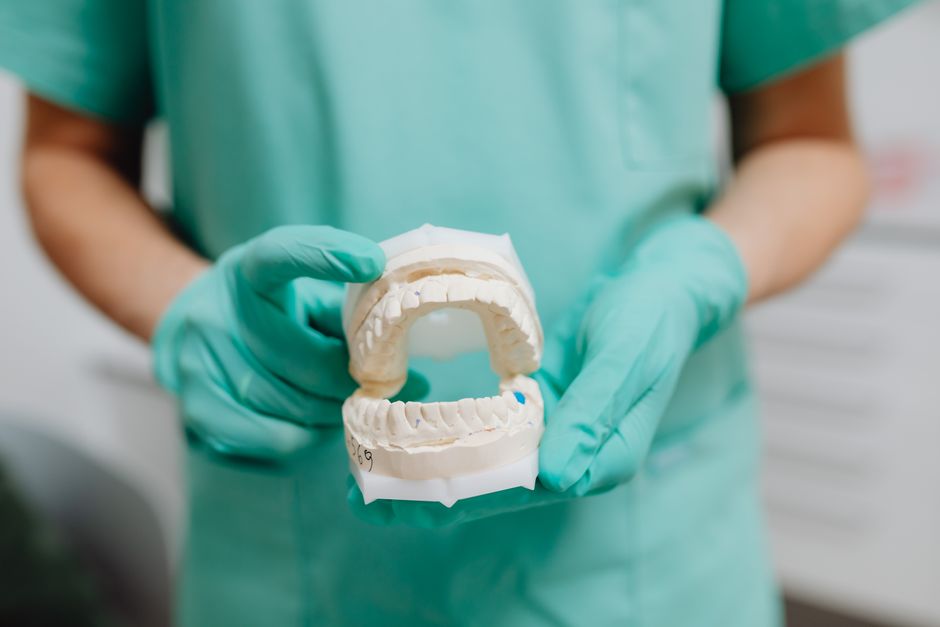
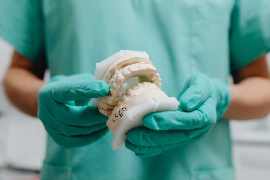
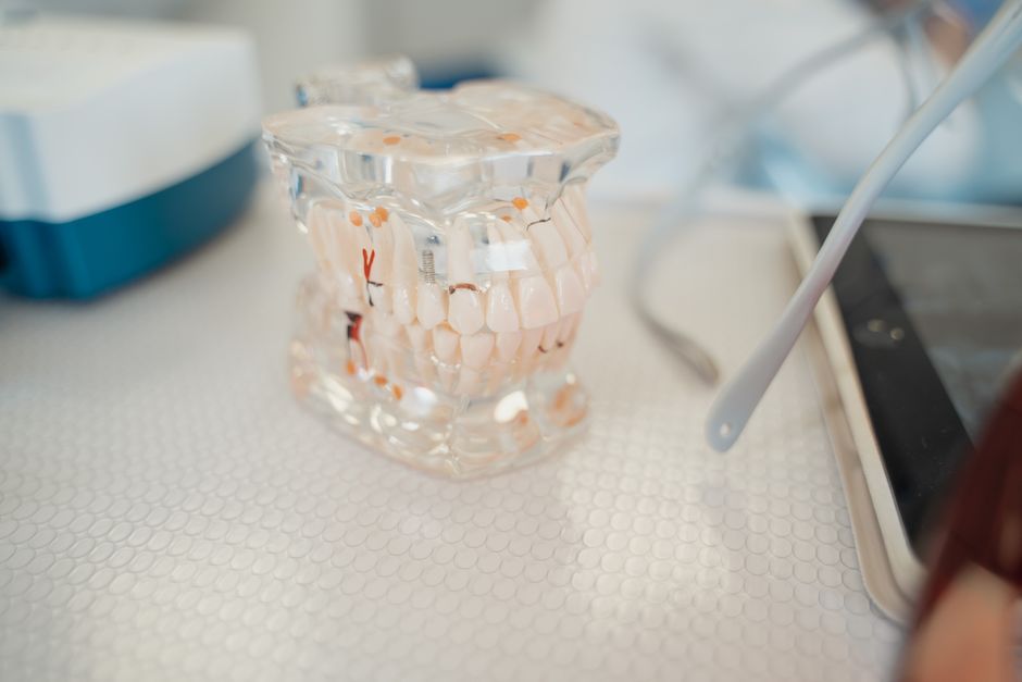
Clinical exam commonly includes palpation of masseter, temporalis, medial pterygoid region (as feasible), assessment of mandibular range of motion, deviation/deflection, joint sounds (with awareness that sounds alone are not necessarily pathology), and inspection for wear facets, linea alba, tongue scalloping, and fractured restorations. Periodontal status should be noted because mobility, inflammation, or attachment loss can change how occlusal forces are distributed.
When periodontal conditions are active, discomfort and occlusal instability can complicate TMD-like symptoms. For a periodontal-inflammation perspective, the clinical mindset used in managing acute conditions such as Necrotizing Ulcerative Gingivitis (NUG): Symptoms, Causes, and Clinical Approach highlights a principle relevant to TMD workups as well: identify the primary driver, control inflammation/pain, and avoid irreversible procedures during unstable phases.
Occlusal Analysis Without Overpromising
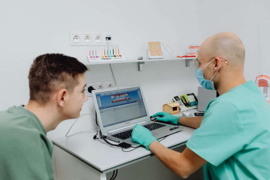
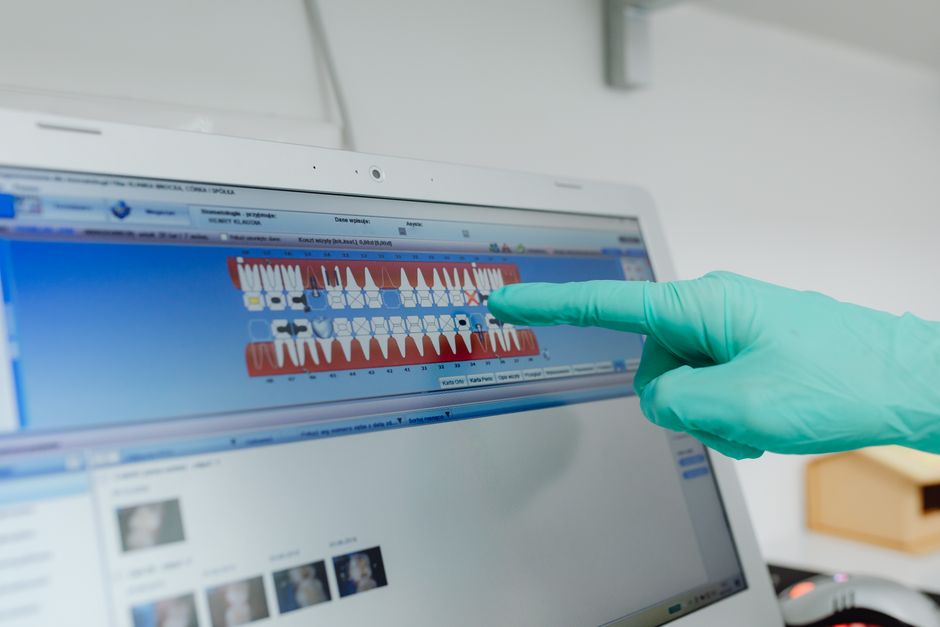
Occlusion can be part of the picture, but it is rarely the only piece. Look for signs of occlusal overload, fremitus, interferences that coincide with symptoms, and changes after recent restorations. Digital dentistry tools—such as intraoral scans and virtual articulation—can document baseline contacts and help communicate findings. However, it is wise to avoid implying that “fixing the bite” will always “cure” TMD. Many TMD presentations are multifactorial, and reversible strategies are typically favored first.
Imaging and Adjunct Tools: When and Why
CBCT, MRI, and the Limits of Imaging
CBCT may be helpful when bony changes are suspected, particularly in degenerative joint disease or pre-surgical planning, while MRI is commonly used to assess soft tissue and disc position. Imaging should be guided by history and exam findings; incidental findings are common, and correlation with symptoms is essential.
Endodontic and Restorative Mimics of TMD Pain
Clinicians regularly encounter toothache-like pain that is actually muscular referral, as well as pulpal pain that patients interpret as “jaw joint” discomfort. Cracked teeth, high restorations, and apical pathology can mimic TMD symptoms. When diagnostic uncertainty exists, magnification can improve confidence in identifying microcracks, accessory canals, or complex anatomy. For example, Dental Operating Microscope in Complex Root Canal Anatomy: Why It Matters reflects how advanced visualization supports differential diagnosis and reduces the risk of misattributing odontogenic pain to TMJ issues.
Management Concepts: Reversible First, Collaborative Always
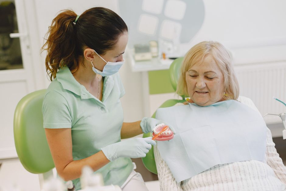
Patient Education and Behavioral Strategies
Education is often the most cost-effective first step. Patients benefit from understanding awake clenching cues, jaw relaxation habits, posture awareness, and the influence of stress and sleep. In many cases, symptom monitoring and habit modification reduce muscle overload. This content is for educational purposes; individualized diagnosis and management should be provided by qualified clinicians.
Occlusal Splints and Protective Approaches
Occlusal appliances are commonly used to protect teeth/restorations and to reduce muscle hyperactivity in selected cases. The clinical focus is typically on reversibility and risk reduction rather than “permanent correction.” Proper design, delivery, and follow-up are essential, especially for patients with mixed muscular and joint signs.
Restorative Dentistry in Bruxers: Material and Design Considerations
When bruxism is present, restorative planning should consider load distribution, guidance concepts, and material selection. Patients requesting esthetic changes may be candidates for conservative approaches (when clinically appropriate), such as direct composite to close spaces. In that context, Advantages of Direct Composite Techniques for Closing Diastema aligns with a minimally invasive philosophy that can be especially relevant in patients with parafunctional risk—where preserving enamel and enabling repairability may be beneficial.
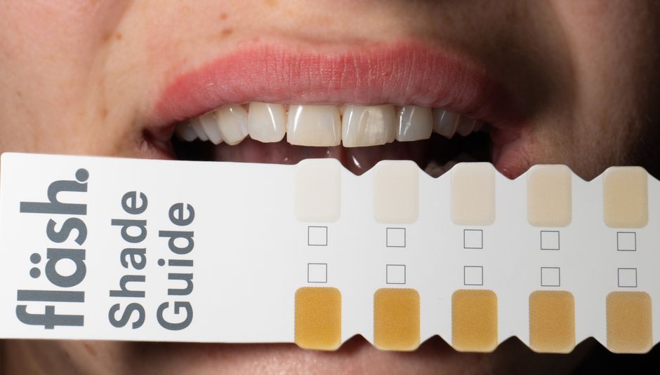
Smile Design, Veneers, and Full-Mouth Planning in the Presence of Bruxism
Why Esthetics Must Be Connected to Function
Patients often pursue veneers or smile makeovers due to chipping, shortening of teeth, or “aged” appearance—features that may be accelerated by bruxism. Yet, aesthetic-only planning can fail if functional risk is not integrated. When increasing vertical dimension, altering guidance, or changing incisal length, clinicians should evaluate muscle comfort, phonetics, envelope of function, and joint tolerance. A structured workflow helps maintain predictability, especially when combining digital planning with physical prototypes.
For clinicians refining their sequence from records to mock-up to definitive restorations, Which Procedures Are Used in Smile Design? A Clinical Workflow for Modern Dentistry provides a useful framework. In bruxism-prone patients, that workflow becomes even more valuable because each step can serve as a checkpoint: Does the patient tolerate the changes? Are guidance and contacts stable? Is there any increase in muscle tenderness?
Porcelain vs. Composite: Practical Risk Conversations
Porcelain restorations can provide excellent esthetics and color stability, but bruxism increases fracture and chipping risk across many restorative materials. Composite options may offer reparability and lower cost, while ceramics may offer surface stability and high esthetics. The decision is case-dependent and should be communicated as risk-managed planning rather than guaranteed longevity.
Implant Dentistry and Bruxism: Managing Occlusal Load
Why Parafunction Matters for Implants
Bruxism is often discussed in implant dentistry because implants lack the periodontal ligament’s proprioception and shock absorption. While the literature varies on how strongly bruxism predicts implant complications, many clinicians consider it a relevant risk factor for mechanical issues such as screw loosening, prosthetic fracture, and wear. Thoughtful occlusal scheme design, prosthetic material choices, and protective appliances may be considered within a comprehensive plan.
Patient systemic factors can also intersect with load management. For example, when planning implants for medically complex individuals, risk assessment becomes multidimensional. The clinical thinking in A Practical Implant Guide for Patients with Well‑Controlled Diabetes illustrates how systemic stability, maintenance, and case selection integrate with local mechanical considerations—an approach equally useful when bruxism is part of the patient profile.
Why Hands-On Training Makes a Difference
From Theory to Chairside Decision-Making
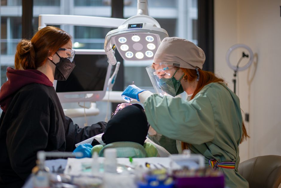
Managing bruxism and TMD-related concerns requires more than memorizing definitions. Clinicians need confidence in differential diagnosis, occlusal records, splint protocols, digital documentation, and restorative design under functional risk. At Istanbul Dental Academy, our continuing dental education approach emphasizes practical workflows—integrating digital dentistry, dental photography for documentation, restorative planning, and prosthodontic principles—so clinicians can apply concepts safely and predictably in real cases.
Skills That Support Better Outcomes
In hands-on settings, dentists can refine core competencies that directly support bruxism/TMD-oriented care: accurate jaw relation records, mock-up evaluation, occlusal adjustment principles, and communication strategies for informed consent. These skills are also highly transferable to implant prosthetics, veneer cases, endodontic diagnosis, and interdisciplinary planning—where misinterpretation of pain sources or occlusal overload can undermine outcomes.
Key Takeaways for Dental Professionals
Bruxism and TMJ disorders frequently overlap, but their relationship is complex and patient-specific. A structured diagnostic process—history, exam, differential diagnosis, and selective imaging—helps clinicians avoid oversimplified conclusions. Reversible, conservative measures are often central early on, while restorative, esthetic, and implant plans should integrate functional risk and long-term maintenance. This content is for educational purposes and is not a substitute for individualized clinical diagnosis or treatment planning.
When clinicians combine evidence-informed concepts with hands-on skill development, they are better positioned to plan predictable restorations, protect patients from avoidable failures, and communicate clearly about risks—especially in bruxism-prone cases where function and esthetics must be planned together.
Diğer Yazılar